Abnormal Shape/Configuration of Corpus Callosum
Susan I. Blaser, MD, FRCPC
DIFFERENTIAL DIAGNOSIS
Common
Normal Variant
Callosal Dysgenesis
Callosotomy
Neoplasm
Lipoma
Glioblastoma Multiforme
Lymphoma, Primary CNS
Decreased White Matter Volume
Hypomyelination
Periventricular Leukomalacia
HIE, Term
Chronic Cerebral Infarction
Diffuse Axonal Injury (DAI)
Multiple Sclerosis
Radiation and Chemotherapy
Obstructive Hydrocephalus
Less Common
Holoprosencephaly
Holoprosencephaly Variants
Rare but Important
Hypertensive Intracranial Hemorrhage
ESSENTIAL INFORMATION
Key Differential Diagnosis Issues
Normal corpus callosum (CC) varies in thickness, shape
Isolated callosal dysgenesis not common
Look for 2nd lesion
Associated CNS anomalies in > 50%
Heterotopia
Cortical dysplasia
Noncallosal midline anomalies
Abnormal brainstem or cerebellum
If not congenital, history crucial!
Helpful Clues for Common Diagnoses
Normal Variant
Size, shape, thickness of normal corpus callosum vary
Splenium, genu are largest parts of corpus callosum
Narrowing between body, splenium (“isthmus”) is normal
Dorsal surface of fully developed, normally myelinated corpus callosum often “wavy”
Immature corpus callosum is thin
Premyelination
Gradually thickens with progressive myelination
Callosal Dysgenesis
One or all segments absent
Rostrum, splenium most likely deficient
Remnants vary in size, shape, configuration
“Micro” corpus callosum
Small, but well-formed
Often syndromic
“Mega” corpus callosum
Isthmus usually absent
Megalencephalic (bulky white matter)
Or small to normal brain (syndromic)
Callosotomy
Surgical disruption
Focal: Approach to 3rd ventricle or suprasellar tumor
Diffuse: Surgery for intractable seizures
Best seen on sagittal or coronal MR
Neoplasm
Can be benign/focal or malignant/diffusely infiltrating
Lipoma
40-50% of interhemispheric fissure
Almost always located in subarachnoid space; blood vessels and cranial nerves course through lipoma; high surgical morbidity → surgery rarely indicated
Common in callosal dysgenesis
Can be bulky, mass-like (“tubonodular” type, usually associated with corpus callosum agenesis; may extend through choroidal fissures into lateral ventricles)
Thin mass curving around corpus callosum body/splenium (“curvilinear” type, corpus callosum present but may be dysgenetic)
Midline lipomas may be part of more general midline developmental disorder
Glioblastoma Multiforme
Most commonly seen in adults, can occur in adolescents (rare)
“Butterfly” glioma
Central necrosis + thick irregular rim enhancement
Lymphoma, Primary CNS
Hyperdense on NECT
Strong, uniform enhancement
Decreased White Matter Volume
Many causes (congenital, acquired)
All may result in focal or diffuse callosal thinning
Hypomyelination
Chromosomal, inborn errors of metabolism
Periventricular Leukomalacia
Premature infant
Increased echogenicity ± loss of normal architecture on ultrasound head
May see cavitation, periventricular cysts
Reduced volume of periventricular white matter
Corpus callosal thinning most commonly in posterior body and splenium
“Scalloped” lateral ventricles
HIE, Term
Term infant with profound partial asphyxia → WM/cortex damaged
Chronic Cerebral Infarction
Axonal loss → focal/diffuse thinning of corpus callosum
Diffuse Axonal Injury (DAI)
20% involve corpus callosum (splenium, undersurface of posterior body)
Multiple Sclerosis
Chronic, late
Obstructive Hydrocephalus
Acute
Corpus callosum stretched, bowed upward
Forniceal columns bowed downward
Chronic
Post-shunt encephalomalacia
Sequela of acute callosal impingement against falx
Helpful Clues for Less Common Diagnoses
Holoprosencephaly
Corpus callosum absent in alobar holoprosencephaly
Large dorsal “cyst” often present
Monoventricle
“Pancake” anterior cerebral tissue
Semilobar may have residual splenium
Frontal fusion and hypoplasia
Caudate head fusion
Splenium may be present
Lobar
Genu may or may not be present
Absent anterior midline falx and fissure
Gray matter often crosses with genu
Holoprosencephaly Variants
Middle interhemispheric variant
a.k.a. syntelencephaly
Splenium, genu present, body deficient
Middle corpus callosum body “dips”
Gray matter crosses at dip
If severe, add bilateral perisylvian polymicrogyria
Helpful Clues for Rare Diagnoses
Hypertensive Intracranial Hemorrhage
Corpus callosum is rare primary site
Image Gallery
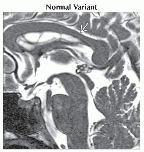 Sagittal T1WI FS MR with a close-up view of the corpus callosum shows normal “wavy” dorsal surface. Note the focal thinning along the posterior body  , a common normal finding. , a common normal finding. |
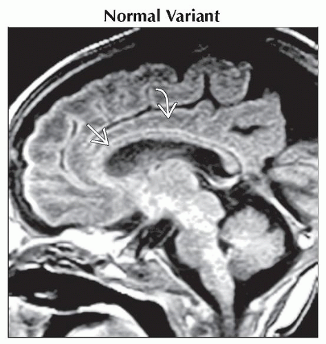 Sagittal T1WI MR shows a normal neonatal corpus callosum  , thin due to age-appropriate lack of myelin maturation. The cingulate gyrus , thin due to age-appropriate lack of myelin maturation. The cingulate gyrus  is normal. is normal. |
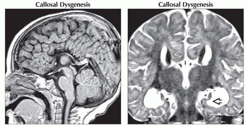 (Left) Sagittal T1WI MR shows callosal agenesis. Note the radial array of paracentral gyri “pointing” to the 3rd ventricle, as well as the absence of identifiable cingulate gyrus. Hippocampal commissure is visualized posteriorly
 . (Right) Coronal T2WI MR shows the absence of crossing callosal fibers, the presence of Probst bundles . (Right) Coronal T2WI MR shows the absence of crossing callosal fibers, the presence of Probst bundles  , and vertical hippocampi , and vertical hippocampi  . .Stay updated, free articles. Join our Telegram channel
Full access? Get Clinical Tree
 Get Clinical Tree app for offline access

|