Ophthalmology Overview
Elias I. Traboulsi
The pediatrician plays an essential role in detecting visual and ocular abnormalities in children. Careful screening of vision by trained and patient assistants and personal examination of the pupils, ocular alignment, red light reflex, and optic nerve head by the pediatrician make it possible to identify such pathologic conditions as major malformations, strabismus, cataracts, and other causes of a white pupil. The child is then referred to the pediatric ophthalmologist for further evaluation and management. Some common ocular problems are easily managed by the pediatrician and referrals are only needed in case of non-resolution. These include nasolacrimal duct obstruction and conjunctivitis. The differentiation of allergic from viral or bacterial conjunctivitis allows the institution of appropriate therapy. In the following brief overview, the basic types of strabismus are covered, and the clinical features, differential diagnosis, and management of some of the common and less common ophthalmic conditions that pediatricians encounter in clinical practice are discussed.
STRABISMUS AND AMBLYOPIA
Ocular misalignment in an infant or child is suspected either because of parental reporting or through direct observation in the office. Pseudostrabismus is the false impression of ocular misalignment as a result of a prominence of epicanthal folds or variations in orbital alignment in a young child. Pseudostrabismus may simulate esotropia (inward deviation of an eye) or, less frequently, exotropia (outward deviation of an eye). Symmetric and centered corneal light reflexes in both eyes and normal fixation patterns are usually sufficient to rule out true strabismus. Parents can be reassured that epicanthal folds will decrease as the child grows and the nasal bridge becomes more prominent, pulling the skin away from the globe and uncovering more of the nasal sclera. A positive family history of strabismus should raise suspicion of true strabismus, in which case a detailed ophthalmologic assessment is always mandatory.
Amblyopia is the loss of vision caused not by an organic ocular or visual pathway lesion, but rather by disuse of one eye and predominant use of the other. The mechanism of vision loss is of visual cortical system origin. This is a reversible process in younger children; in addition to restoring good ocular alignment and binocular vision, one major aim of the treatment of strabismus is the prevention or reversal of amblyopia. Amblyopia therapy consists of penalizing the better-seeing eye to allow stimulation of the central visual centers from the deviated eye. This could be done either by using a patch or by blurring near vision in the better-seeing eye through cycloplegia with atropine drops. Atropine drops are generally not used in very young children. The younger the child, the faster and more dramatic is the response to short periods of occlusion therapy. Longer periods of patching or atropine penalization are required in older children. The upper limit of age at which amblyopia is still reversible is the subject of some debate; it may be approximately 10 years, however some older children may benefit from a trial of penalization.
Congenital or infantile esotropia may not present at birth but is diagnosed in the first 6 months of life. The angle of ocular deviation is usually large, and refractive error is minimal. Associated conditions include overacting inferior oblique muscles and dissociated vertical deviations, which may manifest later in childhood despite initial surgical therapy and good ocular alignment. Surgery should ideally be performed before the age of 1 year, preferably around 6 months, if binocular vision is to be achieved. A positive family history for this likely autosomal-recessive disease with high gene frequency is often elicited.
Accommodative esotropia is one of the most common types of strabismus and becomes evident in the first few
years of life (Fig. 66.1). It is the consequence of accommodative efforts made in response to a relatively large degree of hypermetropia. The dysregulated convergence leads to ocular misalignment and the favoring of one eye with rapid development of amblyopia. Patients should be referred promptly. Therapy consists of the use of corrective glasses and surgery for any residual deviation.
years of life (Fig. 66.1). It is the consequence of accommodative efforts made in response to a relatively large degree of hypermetropia. The dysregulated convergence leads to ocular misalignment and the favoring of one eye with rapid development of amblyopia. Patients should be referred promptly. Therapy consists of the use of corrective glasses and surgery for any residual deviation.
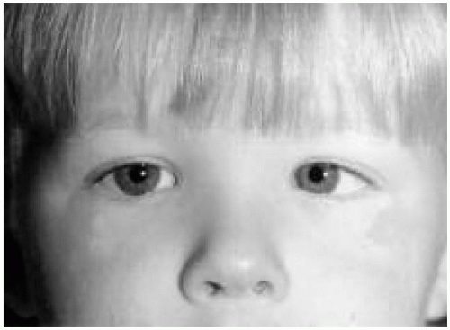 Figure 66.1 Child with left esotropia. Note location of the corneal light reflex at the temporal edge of the pupil. The child also has wide nasal bridge, which accentuates the appearance of esotropia. |
Intermittent exotropia is an intermittent outward deviation of either eye that may become evident when the affected child is tired or ill. Exotropic patients often squint in the sunlight. Treatment consists of the correction of any error of refraction and close follow-up. There is no associated amblyopia. Surgery is indicated if binocular fusion breaks down and the ocular deviation is poorly controlled and is present >50% of the time.
Möbius syndrome is characterized by unilateral or bilateral sixth and seventh nerve palsies. Affected children usually demonstrate esotropia and an expressionless face. Babies with this condition have difficulty breast-feeding and sucking their bottles. Associated anomalies include the Poland anomaly (absence of the pectoralis muscle and radial defects) and terminal limb defects.
Extraocular muscle palsies in children result in incomitant strabismus (different magnitude of the deviation in different directions of gaze), with the largest deviation in the field of action of the affected muscle. Children with acquired palsies may not verbalize a complaint of diplopia, but they may squint, cover one eye with a hand, or assume a compensatory head posture to avoid diplopia.
Third nerve palsies are most commonly caused by trauma or increased intracranial pressure, and they may be complete or incomplete. Other causes include inflammation, infectious and parainfectious processes, vascular lesions, tumors, and degenerative and demyelinating disease involving the nerve. Diabetes is not a cause of third nerve palsy in children. Associated neurologic defects are good clues to the location of the lesion causing the nerve palsy.
Like third nerve palsies, fourth nerve palsies are commonly caused by trauma or tumor, but many are idiopathic and present at birth. An examination of old photographs reveals the characteristic head tilt and provides a good clue to the chronic and benign nature of congenital fourth nerve palsies. Surgery is indicated to relieve torticollis, which may lead to chronic neck pain and possibly back problems.
Sixth nerve palsies may indicate neurologic disease, but many are transitory and benign and follow viral infections. A sixth nerve palsy may be the result of increased intracranial pressure resulting from hydrocephalus, tumor, intracranial hemorrhage, or cerebral edema. It may be caused by trauma, inflammatory conditions such as meningitis, and degenerative or demyelinating conditions. Benign sixth nerve palsy in children develops 1 to 3 weeks after a febrile illness and usually subsides within 6 months. The child with cranial nerve palsy should undergo a complete neurologic evaluation, including computed tomography or magnetic resonance imaging of the head. A history of recent viral disease should be obtained, and the child should receive care from an ophthalmologist and a neurologist. The acute development of nonaccommodative esotropia and weakness of one or both lateral rectus muscles may indicate the presence of a Chiari malformation.
Nystagmus refers to rhythmic oscillations of the eyes that occur independently of normal movements. In pendular nystagmus, the velocity of movement is equal in the two directions. In contrast, jerk nystagmus has slow and fast components. The different kinds of nystagmus are named according to the refixation and the direction in which the nystagmus occurs (e.g., in right-beating jerk nystagmus, the fast refixation component is to the right). In conjugate nystagmus, the binocular oscillations are in phase, whereas disconjugate or dissociated nystagmus can be monocular or binocular with a slow component that is out of phase. Latent nystagmus is elicited by an inter ruption of binocular vision, such as the occlusion of one eye. Infantile nystagmus is present at birth and may be associated with abnormal head movements and positions. Visual acuity is usually decreased. Albinism is probably the most common cause of nystagmus in childhood. Tyrosinase-positive oculocutaneous albinism may be difficult to diagnose except with the slit-lamp. Retroillumination reveals iris transillumination in patients with any type of albinism. In addition, patients with albinism exhibit foveal hypoplasia and misrouting of optic nerve fibers. There are numerous causes of sensory (resulting from poor vision) nystagmus, mostly related to structural ocular abnormalities such as aniridia or cataracts that lead to poor vision. Central nervous disease can lead to
nystagmus through interference with the centers that control eye movement. Nystagmus can be familial and is most commonly inherited in an X-linked fashion.
nystagmus through interference with the centers that control eye movement. Nystagmus can be familial and is most commonly inherited in an X-linked fashion.
OBSTRUCTION OF THE NASOLACRIMAL DUCT
Most cases of obstruction of lacrimal drainage in children are developmental; others are caused by infections, trauma, and lid dysfunction. Obstruction of the nasolacrimal duct, most commonly caused by a failure of the distal membranous end of the nasolacrimal duct to open, occurs in approximately 5% of infants and is bilateral in as many as one third of cases. Obstruction of the nasolacrimal duct may be caused by blockage elsewhere in the lacrimal system or by an absence of the puncti or canaliculi, which interferes with the normal drainage of tears. Rarely, lacrimal obstruction occurs as part of the facial clefting syndromes and the Goldenhar syndrome.
Infants with lacrimal obstruction present with a “weteyed” appearance, persistent or intermittent tearing, and various degrees of mucopurulent discharge over the medial canthal area and lids. Pressure over the area of the lacrimal sac expresses whitish material from the lacrimal puncti. Superimposed dacryocystitis may be present, and dacryocystoceles or fistulae may develop.
Most obstructions (90%) resolve spontaneously by 18 months, and lid hygiene alone is the indicated treatment in most cases. Fingertip compressions over the area of the lacrimal sac, with massage directed centrally while the upper end of the lacrimal system is blocked, may be tried for a short period of time; this maneuver increases pressure inside the system, possibly causing the distal membrane to rupture into the nose. Long-term antibiotic therapy should be avoided. Some pediatric ophthalmologists prefer early probing after a short trial of conservative management for 2 to 4 weeks; this results in early patency of the system and avoids potential infections and continuous irritation and annoyance. Probing is performed in the operating room with the patient under inhalation anesthesia. The surgery is successful >90% of cases. If it fails, it can be repeated with or without silicone intubation of the lacrimal system. Silicone stents are left in place for 3 to 6 months. If probing and silicone intubation fail to maintain a patent system, a dacryocystorhinostomy is performed. This procedure provides direct drainage of tears from the lacrimal sac into the nose. Dacryocystitis should be treated with systemic antibiotics and may resolve only after nasolacrimal probing.
The differential diagnosis of tearing in the infant includes congenital glaucoma. The latter can be differentiated from nasolacrimal duct obstruction by the presence of an enlarged and hazy cornea, sensitivity to light, and elevated intraocular pressure in congenital glaucoma.
LEUKOCORIA (WHITE PUPIL): DIFFERENTIAL DIAGNOSIS OF RETINOBLASTOMA
The differential diagnosis of a white pupillary reflex (or for that matter an abnormal pupillary light reflex) in an infant is the differential diagnosis of retinoblastoma. Leukocoria, or white pupil (Fig. 66.2), is a white or tan reflex in the normally black pupillary area. The reflex, which may be observed in certain ambient lighting conditions or only in certain directions of gaze, can theoretically result from the opacification or tumefaction of any structure behind the iris (e.g., lens, vitreous, retina, choroid). The exact cause of the white reflex should be determined as soon as possible so that treatment of the underlying disease can be started.
Stay updated, free articles. Join our Telegram channel

Full access? Get Clinical Tree


