Chapter 161 Vasculitis Syndromes
Childhood vasculitis encompasses a broad spectrum of diseases that share a common denominator, inflammation of the blood vessels. The pathogenesis of the vasculitides is generally idiopathic; some forms of vasculitis are associated with infectious agents and medications, and others may occur in the setting of preexisting autoimmune disease. The pattern of vessel injury provides insight into the form of vasculitis and serves as a framework to delineate the different vasculitic syndromes. The distribution of vascular injury includes small vessels (capillaries, arterioles, and postcapillary venules), medium vessels (renal arteries, mesenteric vasculature, and coronary arteries), and large vessels (the aorta and its proximal branches). Additionally, some forms of small vessel vasculitis are characterized by the presence of antineutrophil cytoplasmic antibodies (ANCAs), whereas others are associated with immune complex deposition in affected tissues. A combination of clinical features, histologic appearance of involved vessels, and laboratory data is utilized to classify vasculitis (Tables 161-1 to 161-3).
Table 161-1 CLASSIFICATION OF CHILDHOOD VASCULITIS
I. PREDOMINANTLY LARGE VESSEL VASCULITIS
II. PREDOMINANTLY MEDIUM VESSEL VASCULITIS
III. PREDOMINANTLY SMALL VESSEL VASCULITIS
IV. OTHER VASCULITIDES
* Associated with antineutrophil cytoplasmic antibody.
Adapted from Ozen S, Ruperto N, Dillon MJ, et al: EULAR/PReS endorsed consensus criteria for the classification of childhood vasculitides, Ann Rheum Dis 65:936–941, 2006.
Table 161-2 FEATURES THAT SUGGEST A VASCULITIC SYNDROME
CLINICAL FEATURES
LABORATORY FEATURES
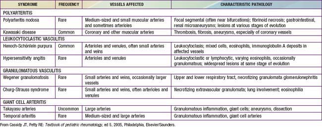
From Cassidy JT, Petty RE: Textbook of pediatric rheumatology, ed 5, Philadelphia, 2005, Elsevier/Saunders.
Childhood vasculitis varies from a relatively benign and self-limited disease such as Henoch-Schönlein purpura to catastrophic disease with end-organ damage as seen in Wegener granulomatosis. Vasculitis generally manifests as a heterogeneous multisystem disease. Although some features, such as purpura, are easily identifiable, others, such as hypertension secondary to renal artery occlusion or glomerulonephritis, can be more subtle. Ultimately, the key to recognizing vasculitis relies heavily on pattern recognition. Demonstration of vessel injury and inflammation on biopsy or vascular imaging is required to confirm a diagnosis of vasculitis.
Bibliography
Dedeoglu F, Sundel RP: Vasculitis in children, Rheum Dis Clin North Am 33:555–583, 2007.
Jennette JC, Falk RJ, Andrassy K, et al: Nomenclature of systemic vasculitides: proposal of an international consensus conference, Arthritis Rheum 37:187–192, 1994.
Jones RB, Tervaert JWC, Hauser T, et al: Rituximab versus cyclophosphamide in ANCA-associated renal vasculitis, N Engl J Med 363(3):211–226, 2010.
Ozen S, Ruperto N, Dillon MJ, et al: EULAR/PReS endorsed consensus criteria for the classification of childhood vasculitides, Ann Rheum Dis 65:936–941, 2006.
Wahezi DM, Gomes WA, Howite NT: Cranial nerve involvement with juvenile polyarteritis nodosa: clinical manifestations and treatment, Pediatrics 126(3):e719–e722, 2010.
161.1 Henoch-Schönlein Purpura
Henoch-Schönlein purpura (HSP) is the most common vasculitis of childhood and is characterized by leukocytoclastic vasculitis and immunoglobulin (Ig) A deposition in the small vessels in the skin, joints, gastrointestinal tract, and kidney.
Epidemiology
HSP occurs worldwide and affects all ethnic groups. The incidence of HSP is estimated at 14-20/100,000 children per year and affects males more than females, with a 1.2-1.8 : 1 male: female ratio. Approximately 90% of HSP cases occur in children, usually between the ages of 3 and 10 yr. HSP is distinctly less common in adults, in whom severe and chronic complications are often encountered. HSP is more common in the fall, winter, or spring and is unusual in summer months. Many cases of HSP follow a documented upper respiratory infection.
Pathology
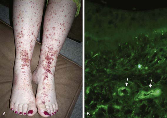
Skin biopsies demonstrate vasculitis of the dermal capillaries and postcapillary venules. The inflammatory infiltrate includes neutrophils and monocytes. Renal histopathology typically shows endocapillary proliferative glomerulonephritis, ranging from a focal segmental process to extensive crescentic involvement. In all tissues, immunofluorescence identifies IgA deposition in walls of small vessels (see Fig. 161-1), accompanied to a lesser extent by deposition of C3, fibrin, and IgM.
Pathogenesis
The exact pathogenesis of HSP remains unknown. Given the frequency of preceding upper respiratory infections, including group A streptococcal infections, an infectious trigger is suspected. The common finding of deposition of IgA, specifically IgA1, suggests that HSP is a disease mediated by IgA and IgA immune complexes. HSP occasionally clusters in families, suggesting a genetic component. HLA-B34 and HLA-DRB1*01 alleles have been linked to HSP nephritis.
Clinical Manifestations
The hallmark of HSP is its rash: palpable purpura starting as pink macules or wheals and developing into petechiae, raised purpura, or larger ecchymoses. Occasionally, bullae and ulcerations develop. The skin lesions are usually symmetric and occur in gravity-dependent areas (lower extremities) or on pressure points (buttocks) (Figs. 161-1 and 161-2). The skin lesions often evolve in groups, typically lasting 3-10 days, and may recur up to 4 mo after initial presentation. Subcutaneous edema localized to the dorsa of hands and feet, periorbital area, lips, scrotum, or scalp is also common.
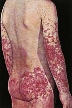
Figure 161-2 Henoch-Schönlein purpura.
(From Korting GW: Hautkrankheiten bei Kindern und Jungendlichen, ed 3, Stuttgart, 1982, FK Schattaur Verlag.)
Musculoskeletal involvement, including arthritis and arthralgias, is common, occurring in up to 75% of children with HSP. The arthritis tends to be self-limited and oligoarticular, with a predilection for the lower extremities, and does not lead to deformities. The arthritis usually resolves within 2 wk but can recur.
Gastrointestinal manifestations of HSP occur in up to 80% of children with HSP. They include abdominal pain, vomiting, diarrhea, paralytic ileus, melena, intussusception, and mesenteric ischemia or perforation. Endoscopic evaluation is usually not needed but may identify purpura of the intestinal tract.
Renal involvement occurs in up to 50% of children with HSP, manifesting as hematuria, proteinuria, hypertension, frank nephritis, nephrotic syndrome, and acute or chronic renal failure. Progression to end-stage renal disease is uncommon in children (1-2%) (see Chapter 509 for more detailed discussion of HSP renal disease).
Diagnosis
The diagnosis of HSP is a clinical one and is often straightforward when the typical rash is present. However, in at least 25% of cases, the rash appears after other manifestations, making early diagnosis challenging. Classification criteria for HSP are summarized in Table 161-4. The differential diagnosis for HSP depends on specific organ involvement but usually includes other small vessel vasculitides, infections, coagulopathies, and other acute intra-abdominal processes.
Table 161-4 CLASSIFICATION CRITERIA FOR HENOCH-SCHÖNLEIN PURPURA*
AMERICAN COLLEGE OF RHEUMATOLOGY CLASSIFICATION CRITERIA†
EUROPEAN LEAGUE AGAINST RHEUMATISM/PEDIATRIC RHEUMATOLOGY EUROPEAN SOCIETY CRITERIA‡
* Classification criteria are developed for use in research and not validated for clinical diagnosis.
† Developed for use in adult and pediatric populations. Adapted from Mills JA, Michel BA, Bloch DA, et al: The American College of Rheumatology 1990 criteria for classification of Henoch-Schonlein purpura, Arthritis Rheum 33:1114–1121, 1990.
‡ Developed for use in pediatric populations only.
Adapted from Ozen S, Ruperto N, Dillon MJ et al: EULAR/PReS endorsed consensus criteria for the classification of childhood vasculitides, Ann Rheum Dis 65:936–941, 2006.
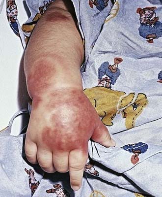
Acute hemorrhagic edema (AHE), an isolated cutaneous leukocytoclastic vasculitis that affects infants <2 yr of age, resembles HSP clinically. AHE manifests as fever; tender edema of the face, scrotum, hands, and feet; and ecchymosis (usually larger than the purpura of HSP) on the face and extremities (Fig. 161-3). The trunk is spared, but petechiae may be seen in mucous membranes. The patient usually appears well except for the rash. The platelet count is normal or elevated, and the urinalysis results are normal. The younger age, the nature of the lesions, absence of other organ involvement, and a biopsy may help distinguish AHE from HSP.
Laboratory Findings
No laboratory finding is diagnostic of HSP. Common but nonspecific findings include leukocytosis, thrombocytosis, mild anemia, and elevations of erythrocyte sedimentation rate (ESR) and C-reactive protein (CRP). Occult blood is frequently found in stool specimens. Autoantibody testing is not useful diagnostically except to exclude other diseases. Serum IgA values are often elevated but are not routinely measured. Assessment of renal involvement with blood pressure, urinalysis, and serum creatinine is necessary.
Ultrasound is often used in the setting of gastrointestinal complaints to look for bowel wall edema or the rare occurrence of an associated intussusception. Barium enema can also be used to both diagnose and treat intussusception. Although often unnecessary in typical HSP, biopsies of skin and kidney can provide important diagnostic information, particularly in atypical or severe cases, and characteristically show IgA deposition in affected tissues.
Treatment
Treatment of HSP is supportive, with an emphasis on assuring adequate hydration, nutrition, and analgesia. Controversy continues concerning the appropriate use of glucocorticoids in the management of HSP, but steroids are most often used to treat significant gastrointestinal involvement or other life-threatening manifestations. Empiric use of prednisone (1 mg/kg/day for 1 to 2 wk, followed by taper) reduces abdominal and joint pain but does not alter overall prognosis nor prevent renal disease. Although few data are available to demonstrate efficacy, intravenous immune globulin and plasma exchange are sometimes used in the setting of severe disease. In some cases, chronic HSP renal disease is managed with a variety of immunosuppressants, including azathioprine, cyclophosphamide, and mycophenolate mofetil. End-stage renal disease develops in up to 8% of children with HSP nephritis.
Complications
Acutely, serious gastrointestinal involvement such as intestinal perforation imparts significant morbidity and mortality. Renal disease is the major long-term complication, occurring in 1-2% of children with HSP. Renal disease can develop up to 6 mo after diagnosis but rarely does so if the initial urinalyses findings are normal. It is recommended that children with HSP undergo serial monitoring of blood pressure and urinalyses for 6 mo after diagnosis, especially those who presented with hypertension or urinary abnormalities.
Prognosis
Overall, the prognosis for childhood HSP is excellent, and most children experience an acute, self-limited course. About 30% of children with HSP experience one or more recurrences, typically within 4-6 mo of diagnosis. With each relapse, symptoms are usually milder than at presentation. Children with a more severe initial course are at higher risk for relapse. Chronic renal disease develops in 1-2% of children with HSP, and approximately 8% of those with HSP nephritis go on to have end-stage renal disease.
Coppo R, Mazzucco G, Cagnoli L, et al. Long term prognosis of Henoch-Schonlein nephritis in adult and children. Italian Group of Renal Collaborative Study on Henoch-Schonlein purpura. Nephrol Dial Transplant. 1997;12:2277-2283.
Davin JC, Weening JJ. Henoch-Schönlein purpura nephritis: an update. Eur J Pediatr. 2001;160:689-695.
Donnithorne KJ, Atkinson P, Hinze CH, et al. Rituximab therapy for severe refractory chronic Henoch-Schönlein purpura. J Pediatr. 2009;155:136-139.
Hoffman GS. Therapeutic interventions for systemic vasculitis. JAMA. 2010;304(21):2413-2414.
Stay updated, free articles. Join our Telegram channel
Full access? Get Clinical Tree
