Urogynecology and Pelvic Floor Dysfunction


Urogynecology and Pelvic Floor Dysfunction


Thomas M. Julian


As the American population ages, the number of women suffering pelvic floor dysfunction increases. These disorders cause changes in normal urination, defecation, sexual function, and pelvic comfort.1


URINARY TRACT INFECTION


Acute Urinary Tract Infection

Acute urinary tract infection (UTI) is a common reason women seek health care. Sexually active women ages 20 to 40 years and postmenopausal women have the highest incidence of acute, uncomplicated UTI. Nearly half of women will experience one UTI in their lifetime, and half of those women will have at least one additional UTI.2 Risk factors associated with UTI are female gender, sexual activity and spermicide-based contraception, and a history of recent UTI. After menopause, UTI may be more common because tissues of the vagina, urethra, and the base of the bladder atrophy and may serve as less of a deterrent to bacterial contamination. Postmenopausal women are also more prone to diabetes and chronic illness(es) that may impair their immune system. Kidney stones and anatomic urinary tract abnormalities (usually diagnosed in childhood) may predispose to infection as will urinary tract catheterization.2,3

Normally, urine is sterile and contains fluids, salts, and waste products. Infection occurs in most cases when fecal bacteria colonize the vaginal introitus and then enter the urethra and multiply. The ureters and bladder normally prevent urine from backing up toward the kidneys, and the flow of urine from the bladder helps wash bacteria out of the body. Glycoproteins line the bladder to help prevent adherence of bacteria to the bladder wall.


Symptoms and Evaluation

In most cases, lower UTI symptoms and signs develop rapidly and may include a strong, persistent urge to urinate accompanied by dysuria, frequency, small-voided volumes, hematuria, cloudy urine, and/or strong-smelling urine. The symptoms of cystitis may be subtle in the very young and the elderly. The probability of cystitis in a woman with any symptom of a UTI is 50%, and if she has dysuria and frequency without any accompanying vaginal discharge or vaginitis, it is 90%.4 Three studies have shown that UTI can be accurately self-diagnosed by women 85 to 95% of the time and that a short-term antibiotic regimen is highly effective curative therapy.5, 6, 7 Symptoms such as malaise, temperature elevation, chills, suprapubic tenderness, flank tenderness, and rigors suggest upper UTI.

Visual inspection of the urine is not helpful in diagnosis other than to identify gross bleeding. Cloudiness is most often due to protein or crystals and malodorous urine from diet or medication. Patients with UTI symptoms should give a clean catch urine specimen for analysis. The higher bacterial count of the first morning void makes it the preferable one for analysis. The urethra is cleansed and urine collected at midstream into a sterile container and sent as soon as possible to be examined. If the specimen will not be sent and processed within 2 hours from collection, it needs to be refrigerated at 4°C and never held for more than 48 hours.

Problems with interpreting a urinalysis because of poor collection technique (contamination) are common in the elderly or disabled and may result in either high false-positive test rates or specimens that cannot be interpreted. In these instances, urine sample collection should be assisted by a nurse or, if necessary, obtained by sterile catheterization.

A fresh specimen can be examined in office with reagent strips (dipstick test) and/or microscopic urinalysis. The dipstick test is simpler and quicker, consisting of dipping a commercially available strip into the fresh specimen. Colormetric reagent boxes on the stick are individual assays for pH, specific gravity, protein, glucose, ketone, bilirubin, urobilinogen, blood, leukocyte esterase (white blood cell [WBC]), and nitrite reductase. After predetermined developing times listed on the bottle containing the strips, the colors of the reagent boxes are compared to references on the bottle. A urine pH greater than or equal to 7.5 suggests UTI. The nitrite test is designed to detect bacteria; it detects greater than 105 colony-forming units (CFU)/mL of Enterobacteriaceae per mL of urine, although it lacks adequate sensitivity for detection of other organisms, so negative results should be interpreted with caution.8 Leukocytes are lysed and liberate esterase when the urine pH is greater than 6.0,
when urine osmolality is low, or when analysis is delayed. Dipstick analysis is most predictive for UTI when the results are positive for bacteria (positive nitrites) and the presence of leukocyte esterase is detected; in these instances, they have a sensitivity of 75% and a specificity of 82% for UTI.4 Some foods may produce false-positive nitrite tests. A negative dipstick, in the presence of significant UTI symptoms, does not rule out an infection and empiric therapy, or further evaluation may be warranted.

Urine microscopy from a centrifuged specimen can be used to diagnose UTI. Bacterial counts are estimated from low-power field (lpf) or high-power field (hpf) counts, and greater than 10 WBC/hpf indicate pyuria. Pyuria is present in almost all women with acute cystitis or pyelonephritis; if it is absent, an alternative diagnosis must be strongly considered.9 The Infectious Disease Society of America has moved away from the traditional threshold of 105 CFU/mL as the cutoff for UTI and now recommends a criterion of greater than or equal to 103 CFU/mL of a typical uropathogen.10

Urinalysis alone is sufficient for the diagnosis of uncomplicated cystitis if the symptoms are consistent with a UTI. Urine culture and sensitivity testing are indicated for failed therapy or suspected antibiotic resistance, recurrent infection, suspected pyelonephritis, uncertain diagnosis after office evaluation, pregnancy, or patients at high risk for UTI.11, 12, 13


Pathology

Quantitative bacterial counts help distinguish infection from contamination, although studies show that lower bacterial counts in symptomatic women can still be important.14 Low-count bacteriuria (102 and 104) with microorganisms typical of UTI may signal an early phase of UTI. Symptoms may start when the urethra is colonized. As a result, some think the microbiologic UTI criterion should be reduced to greater than 102 CFU/mL in symptomatic patients, especially when Enterobacteriaceae are grown. A catheterized urine specimen is considered UTI positive with a growth of 102 CFU/mL. In children, UTI is defined as a bacterial count approximately 104 CFU/mL urine, excluding vaginal contamination of the specimen. For infections with Staphylococcus saprophyticus and Candida species, the lower cutoff is greater than 104 CFU/mL.

Approximately 70 to 90% of UTIs are caused by strains of Escherichia coli. Approximately 5 to 15% of UTIs are caused by Staphylococcus saprophyticus. Other causative organisms include Aerobacter aeruginosa, Klebsiella, Proteus and Pseudomonas, and the more difficult to detect Haemophilus influenzae and Haemophilus parainfluenza, which do not grow well in culture media for enteric bacteria. Other problematic microorganisms include acid-fast bacilli, Campylobacter, Corynebacterium, Legionella, Pneumococcus, Salmonella, Shigella, fungi (Blastomyces and Coccidioides), Neisseria gonorrhoeae, Chlamydia, and Herpes simplex. DNA tests, Gram stain, and acid-fast stains should be performed for those with UTI symptoms, pyuria, and negative culture.15


Asymptomatic Bacteriuria

Asymptomatic bacteriuria refers to isolation of the same organism in bacterial counts greater than or equal to 105 CFU/mL in two consecutive clean catch urine samples in a patient without UTI symptoms.16 Some studies define it as a single positive urine specimen with greater than or equal to 105 CFU/mL, but transient bacteriuria is common in healthy young women, so the requirement for more than one positive specimen for diagnosis may more accurately reflect the true prevalence.17 If the patient is catheterized, asymptomatic bacteriuria is defined as a single catheterized specimen with isolation of a single organism in quantitative counts of greater than 102 CFU/mL.16

The prevalence of asymptomatic bacteriuria increases with age, from about 1% among young girls to greater than 20% among women older than the age of 80 years who live in the community.17,18 Among diabetic women, the prevalence of asymptomatic bacteriuria is 8 to 14% and is related to the duration of disease and presence of long-term complications, and not necessarily any metabolic parameters of diabetes.19

Screening for and treating asymptomatic bacteriuria should be done for pregnant women and for patients who will undergo a urologic procedure where mucosal bleeding is anticipated.16 It is not indicated for the following populations: women (premenopausal, nonpregnant), diabetic patients, the elderly, or patients with spinal cord injury or indwelling urethral catheters because treatment in these cases may contribute to the development of antimicrobial resistance.20 Although women with asymptomatic bacteriuria are at increased risk for UTI, treatment does not reduce the frequency of UTIs or recurrent asymptomatic bacteriuria.17 Although antibiotics may sterilize the urine in cases of asymptomatic bacteriuria, it generally recurs and it is not associated with long-term complications such as hypertension, renal failure, or increased mortality.18,21


Recurrent Urinary Tract Infection

The diagnosis of recurrent UTI is reserved for patients who experience greater than or equal to two UTIs in 6 months or greater than or equal to three within 12 months. Most recurrences are thought to represent reinfection and not just relapse. Most recurrences occur in the first 3 months after the initial infection.24 If the first infection is caused by E. coli, the likelihood of a second UTI developing within 6 months is higher compared to those cases where the first UTI was due to another organism.25 A number of factors appear to be associated with an increased risk for recurrent UTIs.

Prophylactic antibiotic treatment is highly effective in decreasing the risk for recurrent UTI and is recommended for women with two or more symptomatic UTIs within 6 months or three or more within 12 months. Low-dose antibiotics are taken continuously for 6 months or more (see Table 15.2 for prophylactic treatment regimens for women with recurrent UTI). Nitrofurantoin or trimethoprim/sulfamethoxazole (TMPS) once daily offers excellent prophylaxis against E. coli with lesser risk of yeast overgrowth in the vagina with TMPS. Nitrofurantoin may not be used in patients with renal insufficiency and there are some concerns with long-term exposure and associated pulmonary reactions, chronic hepatitis, and neuropathy. Women using fluoroquinolones for prophylaxis must employ effective contraception because these are contraindicated in pregnant women and children. Other adverse events associated with fluoroquinolones include prolonged QTc interval and ruptured Achilles tendons.26,27

Cystitis rarely leads to kidney damage, therefore invasive testing with cystoscopy, intravenous pyelography, tomography, special scans, and renal ultrasound are not usually required prior to starting antibiotic prophylaxis for recurrent UTI. Testing is recommended for children, the elderly, and for those who fail a trial of antibiotic prophylaxis. When recurrences are frequent or a UTI becomes chronic, referral to a urologist or nephrologist for evaluation is recommended.28

Recommended preventive measures for UTI include making sure the patient wipes from front to back after urination or defecation to prevent bacterial contamination, emptying the bladder soon after intercourse, and avoiding chemical irritants to the area such as bubble bath, feminine deodorants, douches, or powders.

Cranberries, blueberries, and lingonberries are fruits containing tannins (proanthocyanidins), which do not treat but may prevent E. coli bacteria from adhering to
cells in the urinary tract, thereby inhibiting infection. These compounds do not prevent infection by urinary acidification as is commonly believed. Although some studies show cranberry juice may help decrease the number of symptomatic UTIs, the data has not definitively demonstrated efficacy, although there is little likelihood of harm in its use for this purpose.29,30 Some research leads to the recommendation of drinking at least one to two cups of cranberry juice daily or taking at least 300 to 400 mg in tablet form twice daily.30


TABLE 15.2 Antimicrobial Prophylaxis Regimens for Women With Recurrent Urinary Tract Infection


Continuous Prophylaxis Regimens


Trimethoprim/sulfamethoxazole


40 mg/200 mg once daily


40 mg/200 mg three times weekly


Trimethoprim


100 mg once daily


Nitrofurantoin


50 mg once daily


100 mg once daily


Cefaclor


250 mg once daily


Cephalexin


125 mg once daily


250 mg once daily


Norfloxacin


200 mg once daily


Ciprofloxacin


125 mg once daily


Postcoital Regimens


Trimethoprim/sulfamethoxazole


40 mg/200 mg or 80 mg/400 mg


Nitrofurantoin


50 mg or 100 mg


Cephalexin


250 mg


Ciprofloxacin


125 mg


Norfloxacin


200 mg


Ofloxacin


100 mg


Probiotics and lactobacilli are beneficial microorganisms that may protect against infections in the genital and urinary tracts. The best-known probiotics are the lactobacilli strains, such as acidophilus, which is found in yogurt, fermented milk products, and dietary supplements. Other probiotics include Bifidobacterium and the lactobacilli rhamnosus, casei, plantarum, bulgaricus, and salivarius, and also Enterococcus faecium and Streptococcus thermophilus. Not all studies show benefit for probiotics in UTI, and the use of these should be considered investigational.31


Acute Nonobstructive Pyelonephritis

Acute uncomplicated pyelonephritis involves the same bacteria as those seen in acute UTI. Pyelonephritis often presents with sudden chills, high fever, and gastrointestinal (GI) upset. In the elderly, it may present as sudden illness, delirium, and/or vascular failure. Pyelonephritis may be managed on an outpatient basis in patients with mild to moderate symptoms who respond well with rehydration and antibiotics over a 12-hour observation period in an outpatient facility. Hospitalization is indicated in instances of severe illness with high fever, pain, debilitation, and an inability to take oral hydration or medications, pregnancy or in instances where adherence to therapy as an outpatient is questionable.

Fluoroquinolones are the only oral antibiotics recommended for the outpatient empirical treatment of acute uncomplicated pyelonephritis.32 In cases where the likelihood of fluoroquinolone resistance is less than 10% (i.e., the community prevalence of resistance is less than 10% and the patient has not travelled to an area with endemic prevalence greater than 10% and the patient has not used a fluoroquinolone in the preceding 3 to 6 months), then empiric therapy such as ciprofloxacin 500 mg orally twice daily for 7 days or 1000 mg extended release once daily for 7 days or levofloxacin 750 mg orally once daily for 5 to 7 days may be started.33,34 This empiric therapy may be started with or without an initial intravenous dose of antibiotics.35,36 If there is hypersensitivity to fluoroquinolones or known resistance, therapy may include TMPS or an oral beta-lactam if the organism is known to be susceptible. If susceptibility data is not available, an initial intravenous dose of a long-acting parenteral antibiotic such as ceftriaxone or a 24-hour period of aminoglycoside use should be given.

Most clinical symptoms of uncomplicated pyelonephritis will respond to antibiotics within 48 hours and, as with uncomplicated cystitis, a follow-up culture is not indicated if symptoms resolve with antimicrobial therapy.


Other Conditions That May Mimic Urinary Tract Infection

Women with UTI symptoms may have another condition causing their symptoms, such as vaginitis, urethritis, interstitial cystitis, dermatitis, or sexually transmitted infections (STIs). Common vaginal infections caused by Trichomonas, Candida, or an anaerobic bacterial milieu may mimic a UTI. Women with painful urination whose urine does not show bacterial growth in culture may have a sexually transmitted urethritis, most commonly Chlamydia trachomatis. Interstitial cystitis is a bladder wall inflammation occurring predominantly in women that may elicit symptoms of a UTI. Disorders in children that mimic UTI include reactions to bubble bath, diaper rash, poor hygiene, and pinworm infestation.


FEMALE URINARY INCONTINENCE

Urinary incontinence (UI), defined by the International Continence Society (ICS), is involuntary loss of urine.37 Most current population-based studies of UI estimate 25 to 45% of women have UI and the incidence increases with age.38 Known risk factors for UI include childbearing, obesity, other urinary symptoms, and functional impairment. UI is common in persons with cognitive impairment, and the presence of cognitive impairment itself is associated with a 1.5- to 3.5-fold increase in the risk of UI.39 The severity of incontinence (frequency and volume) also increases with age.40

UI is common during pregnancy and is estimated to affect 30 to 60% of pregnant women.41 In the postpartum period, 6 to 35% of women report incontinence; about 50% of the women report only mild leakage, and spontaneous resolution is seen in about 70% of cases.41

Women with UI may experience embarrassment, depression, loss of self-esteem, sexual dysfunction, loss of productivity, and social isolation from this problem, but over half may never seek care for their symptoms, even if these occur at least once a week.42,43 Because the prevalence of UI is high among women across the life span and many will not spontaneously bring it up with their providers, women should be specifically screened for UI.44 Often, patients feel UI as a natural part of aging or are unaware that treatments exist, but because incontinence may serve as the signal for a serious, underlying condition such as neurologic disease or even malignancy, it should never be viewed as just an inconvenience of getting older.

Medical morbidities associated with UI include genital candidal infections, cellulitis and/or pressure ulcers, UTIs and urosepsis due to urinary retention or
indwelling catheters, falls and fractures if the woman slips on floor wet with urine, and sleep deprivation if nocturia is present. Other comorbid conditions, medications, and factors influencing patient function may cause or even worsen UI, either transiently or chronically (see Table 15.3 for potential causes or contributors to UI).

The onset and progression of UI, and its attendant psychological stress, is associated with an increased risk of comorbid psychiatric disorders including panic disorder and depression.45,46 The onset of panic disorders is directly proportional to the extent that incontinence alters a patient’s lifestyle, and the incidence of depression is related to the degree of incontinence. The greatest rate of depression is among patients with mixed incontinence.47,48


TABLE 15.3 Identification and Management of Reversible Conditions That Cause or Contribute to Urinary Incontinence


Condition


Management


Affecting the lower urinary tract


Urinary tract infection (symptomatic with frequency, urgency, dysuria, etc.)


Antimicrobial therapy; consider topical estrogen for postmenopausal women with recurrent urinary tract infections.


Atrophic vaginitis/urethritis


Topical estrogen may worsen symptoms of incontinence, although topical estrogen may relieve atrophic vaginitis.


Pregnancy/vaginal delivery/episiotomy


Behavioral intervention; avoid surgical therapy postpartum because condition may be self-limiting.


Stool impaction


Disimpaction; appropriate use of stool softeners, bulk-forming agents, and laxatives if necessary; implement high-fiber intake, adequate mobility, and fluid intake.


Increased urine production


Metabolic (hyperglycemia, hypercalcemia)


Improved control of diabetes mellitus or treatment of hypercalcemia


Excess fluid intake


Reduction in intake of excess fluids, especially diuretic fluids (e.g., caffeinated beverages, including soda and alcohol)


Volume overload


Venous insufficiency with edema


Support stockings, leg elevation, sodium restriction, diuretic therapy


Heart failure


Medical therapy


Impaired ability or willingness to reach a toilet


Delirium


Diagnosis and treatment of underlying causes of acute confusional state


Chronic illness, injury, or restraint that interferes with mobility


Address cause of impaired mobility; remove restraints if possible. Regular toileting; use of toilet substitutes and environmental alterations (e.g., bedside commode, urinal)


Psychological


Appropriate pharmacologic and/or nonpharmacologic treatment


Nocturia


Nocturnal polyuria


Assess evening fluid intake; assess for volume overload; consider sleep apnea if other symptoms, signs, or risk factors present.


Sleep disturbance


Sleep hygiene; assess for pain, depression, and environmental causes; consider sleep apnea if other symptoms, signs, or risk factors present.


From DuBeau CE. Treatment of urinary incontinence. http://www.uptodate.com. Updated October 2011. Accessed December 2, 2013.


The annual direct cost of female UI in the United States is an estimated $12.4 billion, and the majority of those costs are borne by those living in the community ($10.8 billion) compared to women residing in nursing homes ($5.5 billion).49,50 The vast majority of those costs are for routine care (70% of total costs) with only 9% being spent on treatments. Most of the routine costs are borne out of pocket by the women and are not reimbursed by third-party payers.


Incontinence Symptoms of Female Urinary Tract


Urinary Incontinence Symptoms

The International Urogynecology Association (IUGA) and ICS have categorized UI disorders by symptoms37 (Table 15.4).


  • Stress urinary incontinence (SUI) is involuntary UI with effort or physical exertion, such as sneezing, coughing, or activity, although it may occur without provocation if there is significant damage to the urethral sphincter. SUI occurs when an increase in intraadominal pressure overcomes the urethral sphincter mechanisms that keep the sphincter shut; hence, there is a loss of urine in the absence of a bladder contraction. This is the most common cause of UI in younger women and often coexists with urge incontinence in middle-aged or older women.51


  • Urgency urinary incontinence (UUI) is the involuntary loss of urine associated with a strong urge to void, even when the bladder is not full. Its etiology is presumed to be uninhibited bladder contractions, also referred to as detrusor overactivity.52 This is most likely not the only etiology, however, because detrusor overactivity may be found in continent individuals as well.


    TABLE 15.4 Differential Diagnosis of Urinary Incontinence


    Stress urinary incontinence


    Hypermobility (anatomic)


    Intrinsic sphincter deficiency


    Overactive bladder


    Motor urge incontinence


    Sensory urge incontinence


    Detrusor dyssynergia


    Detrusor hyperreflexia


    Detrusor hyperactivity with incomplete contractility


    Mixed incontinence


    Bypass of the continence mechanism


    Fistula


    Diverticulum


    Ectopic ureter


    Overflow incontinence


    Functional


  • Mixed urinary incontinence (MUI) is involuntary UI associated with urgency and also with physical exertion; it is believed to occur when there is coexisting urgency and stress incontinence.


  • Nocturnal enuresis is involuntary urine loss during sleep.


  • Continuous UI is continuous involuntary urine loss and may be seen with either bladder outlet obstruction and/or impaired detrusor contractility. Patients may be unaware of the leakage at all. Although referred to as “overflow incontinence” in the past, the preferred term mentioned earlier is best to use along with any known associated pathophysiology.


  • Postural UI is involuntary loss of urine associated with change of body position (rising from a seated or lying position).


  • Insensible UI is when UI occurs but the woman is unaware of how it occurred.


  • Coital UI is UI with coitus, occurring with penetration or intromission or UI occurring at orgasm.


Bladder Storage Symptoms

Bladder storage symptoms that may or may not be associated with UI include more frequent urinations during waking hours than before; although seven episodes of voiding during waking hours is often considered the upper limit of normal, it may be higher in some populations. Other symptoms related to bladder storage issues include nocturia, the interruption of sleep one or more times a night to urinate, or a significant and sudden urgency to void.37


Sensory Symptoms

Sensory symptoms encompass a departure from normal sensation(s) or function during bladder filling. Most individuals are aware of their bladders filling up, ultimately resulting in a strong desire to void. With increased bladder sensation, the patient is able to postpone voiding, unlike the urgency sensation associated with UUI where the patient is not able to postpone voiding. Other situations may involve a reduced bladder sensation, that is, the patient is aware the bladder is filling but the strong desire to void does not occur until later than it had in the past. Some patients may complain of absent bladder sensation and are both unaware of the bladder filling and lack any definite desire to void.37


Voiding Symptoms

Voiding symptoms are any departure from normal sensation or function experienced by the woman during or following urination. These include symptoms of hesitancy, slow stream, intermittent flow, straining to void, spraying or splitting of the urinary stream, incomplete bladder emptying, immediate need to void again after passing urine, needing to assume a specific position to void spontaneously or improve bladder emptying, dysuria, and urinary retention despite persistent effort to void.37


Initial Evaluation of Female Urinary Incontinence


Complete History and Physical Examination

The history should focus on the onset and course of incontinence and any associated symptoms. Information regarding how frequently the incontinence occurs, the amount of urine lost, the timing of the incontinence, and precipitants to the incontinence should be noted. The patient should be asked about her bowel and sexual function, the status of any other medical conditions and symptoms she may have, the use of all medications (including over the counter), and any previous surgeries, including any treatment(s) for incontinence and the outcome. A dietary review, including liquids, should be done (Table 15.5).

In general, urgency symptoms are sensitive and specific for the diagnosis of urge incontinence and a report of such symptoms is a fairly reliable diagnostic tool.53,54 Symptoms associated with stress are fairly reliable but are not very specific in identifying SUI because they may also occur with detrusor overactivity or incomplete bladder emptying.53

Symptoms of slow, hesitant, or interrupted voiding patterns, straining to void, nocturia, and frequency are not diagnostically specific. Nocturia may be due to nocturnal polyuria, sleep disturbances, or a lower urinary tract issue such as detrusor overactivity.

Patients with incontinence should optimally have a comprehensive physical examination. The cardiovascular examination should note if there is any evidence of fluid overload. The abdomen should be examined for any masses or tenderness. The extremities should be examined for mobility, function, and evidence of peripheral edema.

The pelvic examination should include a close inspection of the genitalia and vaginal vault for signs of atrophy, stenosis, or inflammation. The urethra is examined for diverticulum or fistula. Urethra and bladder palpation is done to assess for a mass or tenderness. Pelvic floor muscle function is assessed visually and digitally including tone, muscle strength (static and dynamic), voluntary muscle relaxation (absent, partial, complete), muscular endurance (ability to sustain maximal or near maximal force), repeatability (the number of times a contraction to maximal or near maximal force can be performed), duration, coordination, displacement, and configuration. Assessment is bilateral, looking for unilateral defects and asymmetry.55 Urethral hypermobility may be visually assessed by asking the woman to perform a Valsalva maneuver, although it may be hard to determine due to body habitus or a large cystocele. In these cases, urethral hypermobility can be evaluated by placing a cotton-tipped woodentailed applicator, lubricated with sterile 2% lidocaine
gel, into the cleansed urethral meatus to the level of the urethrovesical junction (UVJ) and asking the patient to strain (Q-tip test). The straining angle of the arm of the tail compared with the horizontal axis is the measurement of urethral or bladder neck mobility. A straining angle greater than 30 degrees is indicative of poor anatomic support of the UVJ and urethral hypermobility but does not necessarily confirm the diagnosis of SUI.


TABLE 15.5 Medications That May Cause or Worsen Urinary Incontinence


Medication


Effect on Continence


Alcohol


Frequency, urgency, sedation, delirium, immobility


α-Adrenergic agonists


Outlet obstruction (men)


α-Adrenergic blockers


Stress leakage (women)


Angiotensin-converting enzyme inhibitors


Associated cough worsens stress and possibly urge leakage in persons with impaired sphincter function.


Anticholinergics


Impaired emptying, retention, delirium, sedation, constipation, fecal impaction


Antipsychotics


Anticholinergic effects plus rigidity and immobility


Calcium channel blockers


Impaired detrusor contractility and retention; the dihydropyridine agents can cause pedal edema, leading to nocturnal polyuria.


Estrogen


Worsens stress and mixed leakage in women


GABAnergic agents (gabapentin, pregabalin)


Pedal edema causing nocturia and nighttime incontinence


Latanoprost


Urge incontinence


Loop diuretics


Polyuria, frequency, urgency


Narcotic analgesics


Urinary retention, fecal impaction, sedation, delirium


Nonsteroidal anti-inflammatory drugs


Pedal edema causing nocturnal polyuria


Oral contraceptivesa


Stress, urge, and mixed incontinence


Sedative hypnotics


Sedation, delirium, immobility


Thiazolidinediones


Pedal edema causing nocturnal polyuria


Tricyclic antidepressants


Anticholinergic effects, sedation


Cholinesterase inhibitors


Alone may increase incontinence; increased functional impairment when combined with anti-incontinence antimuscarinic agents


β-Blockers


Urge incontinence


Opioid analgesics


Sedation, anticholinergic effects


H1-receptor antagonists


Confusion


Lithium


Polyuria


GABA, gamma-aminobutyric acid.


a Townsend MK, Curhan GC, Resnick NM, et al. Oral contraceptive use and incident urinary incontinence in premenopausal women. J Urol. 2009;181:2170.


Reprinted with permission from DuBeau CE. Urinary incontinence. In: Pompei P, Murphy JB, eds. Geriatrics Review Syllabus: A Core Curriculum in Geriatric Medicine. 6th ed. New York: American Geriatrics Society; 2006:185. Table based on data from Resnick NM. Geriatric medicine. In: Isselbacher JK, Braunwald E, Wilson JD, et al, eds. Principles of Internal Medicine. New York: McGraw-Hill; 1994:34.


The pelvic support system should be assessed by a split-speculum exam: Remove the top blade of the speculum and hold the bottom blade firmly against the posterior vagina while asking the woman to cough (or perform a Valsalva maneuver) and look for evidence of urethral hypermobility or an anterior wall support defect during the cough. Although loss of any urine associated with these efforts is noted as a positive cough stress test and is helpful in confirming stress leakage, a negative test is less useful because it may occur from patient inhibition or an insufficient amount of urine in the bladder.53 The posterior blade should then be placed anteriorly and held firmly while the posterior vaginal wall is inspected during a cough (or Valsalva maneuver) for evidence of loss of wall support. Bimanual examination should be performed to detect coexistent adnexal or uterine pathology.

Patients should be screened for depression and their cognitive function, as well as functional status, should be noted. The neurologic exam should assess gait, balance, and neurologic signs or symptoms indicative of cervical spondylosis or stenosis, Parkinson disease, multiple sclerosis, peripheral neuropathy, and other neurologic diseases. Vibration and peripheral sensation should be assessed for peripheral neuropathy. The T10 to S4 nerve roots are primarily responsible for voiding control, and neurologic screening should assess lumbosacral spinal segments, including asymmetry of large muscle groups or deformity of the extremities as indirect evidence of bladder function. Motor function can be assessed by flexion and extension maneuvers against resistance at the ankle, knee, and hip. Normal sensation in the upper leg and perineal dermatomes helps confirm intact sensory innervation of the lower urinary tract (Figs. 15.1 and 15.2). Patients with extensive osteoarthritis may suffer from cervical spondylosis or stenosis, which may result in
detrusor overactivity; examination of such patients should include assessment of lateral rotation and flexion of the neck, inspection of the hands for interosseous muscle wasting, and determination if a Babinski reflex is present because these would suggest cervical changes and resultant nerve impairment.


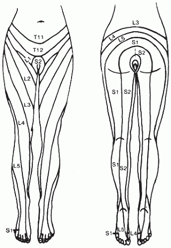
FIGURE 15.1 Dermatome map of lower extremities. (From Ostergard DR, Bent AK. Urogynecology and Urodynamics: Theory and Practice. 4th ed. Baltimore: Lippincott Williams & Wilkins; 1996:682, with permission.)


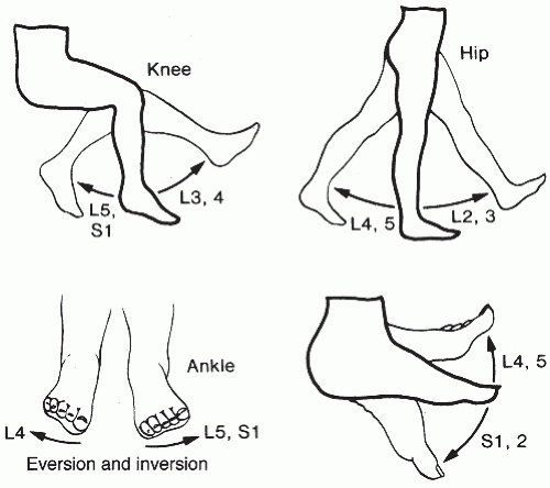
FIGURE 15.2 Testing of motor strength. (From Ostergard DR, Bent AK. Urogynecology and Urodynamics: Theory and Practice. 4th ed. Baltimore: Lippincott Williams & Wilkins; 1996:682, with permission.)

Neurologic evaluation of the genital/pelvic area should include an evaluation of perineal sensation and checking for reflex contraction of the pelvic floor in response to light stroking of the perineal skin lateral to the anus (anal wink) or clitoris (bulbocavernosus reflex); positive responses provide evidence of the integrity of the sacral reflex center. Rectal exam should assess sphincter tone and rule out fecal impaction, which is often associated with UI in the elderly. Vibration and peripheral sensation should be evaluated as a means of assessing for peripheral neuropathy. Findings suggestive of neurologic deficits should be referred for formal neurologic evaluation.


Noninvasive Evaluations of Urinary Incontinence

A urinalysis should be performed for all patients complaining of UI or other symptoms. In older patients, results must be interpreted carefully because there is a high prevalence of asymptomatic bacteriuria in this population; this is not a cause of incontinence.

Pelvic floor questionnaires evaluate the impact of pelvic floor disorders on urinary, bowel, comfort, and sexual function and may help determine the magnitude of the problem for the patient in a way that helps the provider. Having the patient identify the aspect of her incontinence that causes her the most difficulty, either spontaneously or through a questionnaire, may help to target or possibly prioritize treatment approaches. In the United States, some of the questionnaires commonly used include Pelvic Organ Prolapse/Urinary Incontinence Sexual Questionnaire (PISQ-12), Incontinence Impact Questionnaire (IIQ), Urogenital Distress Inventory (UDI), and the Overactive Bladder Treatment Satisfaction Questionnaire (OAB-S).56 The 3IQ test has been developed as a simple means of distinguishing between stress and urge incontinence (Fig. 15.3).57

Asking the patient to keep a bladder diary, or urolog, may be clinically helpful, especially for issues of nocturia, high urinary frequency and/or incontinence frequency, or when the history is unclear. Patients are given a urolog and instructions on what information needs to be recorded as well as how to record the information and a measuring container that fits over the toilet bowl (Fig. 15.4). She records the time and volume/type of her fluid intake and spontaneous voids over a 24- to 72-hour time period (this does not have to be consecutive) while also recording her activities. Additional information regarding prevoid urgency, incontinent episodes (and estimated amounts, e.g., drops, small, medium, and soaking), activity precipitating incontinence, pad usage, episodes of urgency, and abnormal sensation may also be recorded. Although reliability is greatest with the use of 7-day diaries, these can be difficult to keep so it is quite common to use 2- or 3-day diaries in both clinical and research settings.58 Typically, eight voids or fewer during the day are considered normal, and nocturnal frequency should be no more than two times during the night. Polydipsia should be considered when fluid intake exceeds 40 mL/kg body weight during 24 hours, and nocturnal polyuria occurs when over 20 to 30% of total daily fluid volume ingested is voided at night.

Quantification of the amount of urine lost during incontinence episodes may be done by measuring the increase in the weight of perineal pads that are weighed pre- and post-testing. This gives a guide to the severity of incontinence. The pads may be worn over a short period such as 1-hour or 1- to 2-day test periods, and the patient may partake in a variety of activities ranging from normal everyday ones to provocation with defined stress regimens. The perineal pad test, with or without a urinary dye, also is useful to assess urine loss when the patient does not know whether the fluid being lost is urine. The patient wears the pad, and the pad is observed (when dye is used to color urine) or weighed.


Urologic Testing

Although one often sees the word “urodynamics” to describe this testing, there is no predetermined, all-inclusive combination of tests that is described by this term. Urodynamics testing is based on the premise that any situation where intravesical pressure is higher than
urethral closure pressure will result in urinary leakage, and this can be due to either decreases in urethra closure pressure caused by urethral dysfunction or increases in bladder pressure caused by detrusor dysfunction or a combination of both. There are a multitude of clinical tests measuring various properties of the lower urinary tract that have been developed—from simple office cystometrics to multichannel video studies.37,59 For many, urodynamic testing has become routine practice, but its role in the evaluation of incontinence for all is controversial. It is expensive, requires specialized equipment and training, does not always correlate with symptoms, and is invasive (Table 15.6).60,61 Although it is associated with about a 2% risk of UTI, prophylactic antibiotics are not necessary.62 Studies trying to demonstrate better treatment outcomes in instances of UI based on urodynamic testing results compared to treatment based on history and physical examination alone have been mixed.63,64 One meta-analysis that included 1000 women with clinical symptoms of stress incontinence showed that when compared against a urodynamic diagnosis, testing was 91% sensitive but only 51% specific in diagnosing pure stress incontinence. The same analysis showed that for urge
incontinence, history alone was 73% sensitive but only 55% specific.65 For these reasons, clinicians should be aware of what diagnostic information urodynamic tests can and cannot provide, its limitations and issues, and how to accurately interpret or even studies that seek to establish their efficacy.66


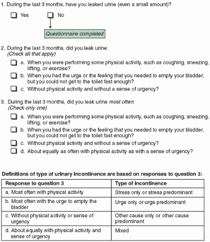
FIGURE 15.3 The 3 incontinence questions. (Reproduced with permission from Brown JS, Bradley CS, Subak LL, et al. The sensitivity and specificity of a simple test to distinguish between urge and stress urinary incontinence. Ann Intern Med. 2006;144:715. Copyright © 2006 American College of Physicians.)


FIGURE 15.4 Example of urolog. (From DuBeau CE. Treatment of urinary incontinence. http://www.uptodate.com. Updated October 2011. Accessed December 2, 2013.)


TABLE 15.6 Indications for Urodynamic Testing


  • History is unclear and/or the diagnosis is uncertain.


  • Invasive or surgical treatments are being considered for treatment.


  • Conservative management has failed.


  • Mixed stress and urge symptoms are present.


  • History of prior surgery for incontinence or prolapse


  • Neurologic disorders are present or suspected.


Controversy exists with respect to the indications for urodynamic testing prior to conservative therapy for overactive bladder (OAB). From a practical standpoint, if the symptoms of urge and frequency dominate the patient history, a postvoid residual (PVR) in some instances, (discussed later) and making sure the patient
does not have a medical contraindication would seem to be all that would be necessary to initiate therapy. There are no randomized controlled trials showing prospective urodynamic testing improves treatment outcomes for either SUI or OAB.67, 68, 69 The testing is also invasive, may be embarrassing for patients, and not cost-effective to apply as a universal policy. Experts mainly agree it is not necessary to perform urodynamic testing on patients prior to instituting conservative management.70

The most commonly performed urodynamic study is filling and voiding fluid cystometry, a test that is also referred to as a cystometrogram (CMG). The term is also used to indicate the graphic results of the measurements made during cystometrography. Fluid cystometry provides information on bladder proprioception (sensation) and capacity, may demonstrate the presence of detrusor overactivity (uninhibited contractions), and evaluates bladder contractility and voiding efficacy. Carbon dioxide was used in the past for cystometry, but it is unreliable because the gas is compressible, irritates the bladder, and does not allow for stress testing or any voiding studies. The use of simple office cystometry, single-channel or multichannel cystometry, ambulatory urodynamics, or videourodynamics is not necessary before starting conservative treatment for UI, or for the women with a clear clinical diagnosis of SUI; nor is it recommended for the patient who seems to have straightforward OAB symptoms.

For those patients undergoing CMG evaluation, the patient comes to the testing visit with a comfortably full bladder. Some urogynecologists advise that the patient empty her bladder and then have any PVR measured. This may be done with a bladder scan, pelvic ultrasound, or directly with a catheter. Ultrasound allows immediate measurement but is less accurate (85 to 94% accuracy) than using a catheter for PVR measurement.71 A PVR less than 50 mL is normal when total bladder volume is 150 mL or more, whereas a PVR greater than 200 mL suggests urinary retention or poor detrusor contractility. The clinical significance of volumes between 50 and 200 mL is unclear. However, the recommendation of measuring PVR is based on opinion and is not supported by any high-quality evidence from randomized trials. The 4th International Consultation on Incontinence recommends against PVR testing as part of the initial evaluation.60 A single PVR requires later repeat confirmation before being considered significant37 (Table 15.7).

CMG testing is a pressure and volume recording during bladder filling. Cystometry can be done with only one channel measuring bladder pressure alone, although the information obtained with single-channel measurements may not be highly reproducible. Use of an additional channel that simultaneously measures abdominal pressure through the rectum or vagina is preferred. Use of multichannel testing can differentiate between changes in abdominal versus bladder pressure changes, that is, the increases in pressure are due to a detrusor contraction and not simply tensing of the abdominal wall. Uninhibited bladder contractions, which represent detrusor overactivity, are present in 8 to 63% of UI patients. The incidence of unstable contractions is increased by making filling CMG more provocative (e.g., faster filling speed, change in posture from supine to standing, cold-water filling, repetitive coughing, heel bouncing, hand washing). Some equipment will also allow for measurement of urethral pressure, which is discussed later in the chapter as urethral leak point pressure. The most common reason to do CMG testing is to distinguish between stress incontinence and detrusor overactivity; CMG testing may also help identify patients with mixed incontinence or abnormalities with bladder sensation.


TABLE 15.7 Patients in Whom Postvoid Residual Measurement May Be Helpful


  • A recurrence of incontinence or development of a new type of incontinence after anti-incontinence surgery


  • Significant pelvic organ prolapse


  • Presence of specific neurologic disease (e.g., spinal cord injury, Parkinson disease)


  • Failed empiric antimuscarinic drug therapy


  • History of recurrent urinary tract infections


  • Urodynamic testing shows detrusor underactivity or bladder outlet obstruction


  • History of urinary retention


  • History of severe constipation


  • Use of medications known to possibly suppress detrusor contractility or increase sphincter tone


  • History of diabetes mellitus with peripheral neuropathy


There are tests called “eye ball” or “poor man” office cystometrics where a catheter attached to a graduated measuring device is used to fill the bladder while observing changes in the fluid level (thought to represent changes in bladder pressure) visually through the transparent measuring device. This approach supposedly assesses detrusor activity and bladder sensation, capacity, and compliance. However, because there are no transducers to demonstrate the source of changes in pressure, it is not possible to distinguish between increased abdominal tension or a detrusor contraction. It is highly possible that use of this methodology will lead to overestimation of detrusor activity. This being the case, it would seem a poor choice to rely on the “eye ball” technique to detect these subtle differences.37,72

Presently, the preferred method of cystometric evaluation is called complex multichannel CMG. These studies are generally performed in a urodynamic laboratory and involve bladder filling with a specified liquid at body temperature (usually water or saline) at a selected rate. Most clinicians fill the bladder at a rate of 50 to 100 mL/minute (faster rates may provoke detrusor activity). The pressure recording system is calibrated to zero at the superior edge of the pubic symphysis. To start, the bladder is empty. Patients are placed in a sitting or semistanding rather than the supine position.


During filling CMG, several data points are obtained. These include the volume at which the patient experiences the following:


  • The first sensation of bladder filling


  • A desire to void but one that can be tolerated


  • Urgent need to void


  • Maximum capacity; patient can no longer avoid micturition

During testing, any abnormal bladder contractions— seen as increases in detrusor pressure—whether they are spontaneous or provoked are noted and are abnormal. At the end of testing, the volume infused into the bladder divided by the change in detrusor pressure is calculated to give an estimate of bladder compliance. Bladder compliance is expressed as mL per cm H2O and can be affected by bladder filling; faster filling is more provocative. Reduced or absent bladder sensation during CMG is noted. Pain during filling CMG is abnormal, and if it occurs, its site, character, and duration is noted.

There are no recognized standards for normal values on CMG. Commonly used ranges include 100 to 200 mL at the first desire to void, a normal desire to void is often felt between 150 and 350 mL, urgency may be noted between 250 and 500 mL, and maximum cystometric capacity (when patient can no longer delay micturition) ranges between 300 and 600 mL. A small rise in detrusor pressure (i.e., less than 10 to 15 cm H2O) without undue sensation of urgency up to a capacity of 400 to 500 is also normal. These values are reflective of wide ranges of normal, and testing with results outside these values is not conclusive for pathology per se. Correlation between symptoms, physical findings, and testing results must be done to arrive at a meaningful assessment (Table 15.8).

Detrusor activity (inherent bladder wall pressure) should change very little with bladder filling. Detrusor overactivity is the occurrence of involuntary detrusor contractions during filling CMG and is seen as wave forms on the CMG of variable duration and amplitude. Symptoms, such as urgency and/or urgency incontinence, may or may not occur. If a neurologic disease is the cause for contractions, then neurogenic detrusor overactivity is diagnosed; otherwise, idiopathic detrusor overactivity is the term used.


TABLE 15.8 Known Issues Affecting Urodynamic Testing Results and/or Interpretation of Results or Studies


  • Urodynamic testing lacks standardization of technical details such as patient position, type of pressure sensor, and filling rate; each of these variables significantly affect results.


  • The setting of a urodynamic laboratory is known to nonphysiologic results in some patients.


  • Use of a transurethral catheter may unmask stress incontinence in some patients.


  • There may be inconsistency in the reproducibility of test results in the same patient.


  • There is a wide range of physiologic values in normal, asymptomatic patients.


The absence of a specific abnormality during urodynamic testing does not conclusively show it does not exist; conversely, not all abnormalities found during urodynamic testing are clinically significant.

Only gold members can continue reading. Log In or Register to continue

Stay updated, free articles. Join our Telegram channel

Jun 25, 2016 | Posted by in GYNECOLOGY | Comments Off on Urogynecology and Pelvic Floor Dysfunction

Full access? Get Clinical Tree

Get Clinical Tree app for offline access