Chapter 541 Urinary Lithiasis
Urinary lithiasis in children is less common in the USA than in other parts of the world. The wide geographic variation in the incidence of lithiasis in childhood is related to climatic, dietary, and socioeconomic factors. Approximately 7% of urinary calculi occur in children <16 yr of age. In the USA, many children with stone disease have a metabolic abnormality. The exceptions are patients with a neuropathic bladder (Chapter 536), who are prone to infection-initiated renal stones, and those who have urinary tract reconstruction with small or large intestine, which predisposes to bladder calculi. The incidence of metabolic stones is similar in boys and girls; they are most common in southeastern USA and are rare in African-Americans. In Southeast Asia, urinary calculi are endemic and are related to dietary factors. Contamination of formula with the organic base and unethical nitrogen-containing food additive melamine has been reported in China.
Stone Formation
Approximately 75% of all stones contain calcium as a major constituent, and 60% are composed of calcium oxalate. Most “spontaneous” stones are composed of calcium, oxalate, or phosphate crystals; others are due to uric acid, cystine, ammonium crystals, or phosphate crystals, or a combination of these substances (Table 541-1). The risk of stone formation increases in the presence of increasing concentrations of these crystals and is reduced with increasing concentrations of inhibitors. Renal calculi develop from crystals that form on the calyx and aggregate to form a calculus. Bladder calculi may be stones that formed in the kidney and traveled down the ureter, or they can form primarily in the bladder.
Table 541-1 CLASSIFICATION OF UROLITHIASIS
CALCIUM STONES (CALCIUM OXALATE AND CALCIUM PHOSPHATE)*
CYSTINE STONES
Cystinuria
STRUVITE STONES (MAGNESIUM AMMONIUM PHOSPHATE)
URIC ACID STONES
INDINAVIR STONES
MELAMINE
NEPHROCALCINOSIS
ACTH, adrenocorticotropic hormone.
Diagnosis
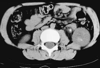
In a child with suspected renal colic, there are multiple imaging options. The most accurate study is an unenhanced spiral CT scan of the abdomen and pelvis (Fig. 541-1). This study takes only a few minutes to perform, has 96% sensitivity and specificity in delineating the number and location of calculi, and demonstrates whether the involved kidney is hydronephrotic. However, the radiation exposure is high. An alternative is to obtain a plain radiograph of the abdomen and pelvis plus a renal ultrasonogram. These studies can demonstrate hydronephrosis and possibly the calculus on the radiograph; however, the calculus is not visualized on sonography unless it is adjacent to the bladder. Consequently, the clinician needs to carefully balance the risks of CT imaging against the lower sensitivity of the plain abdominal film plus sonography.
In 2008 the Society for Pediatric Radiology initiated the Imaging Gently initiative to educate providers on the risks of radiologic imaging in children and to encourage the use of limited imaging in children, particularly those with suspected urolithiasis (http://www.pedrad.org/associations/5364/ig/). This approach was also advocated by the National Quality Forum. In a child with an already-diagnosed calculus, serial plain x-rays or renal ultrasonography can be used to follow the status of the calculus, such as whether it has grown or diminished in size or has moved. If a child has a renal pelvic calculus, a ureteropelvic junction obstruction should be suspected. In some cases, it can be difficult to determine whether hydronephrosis in such a child is secondary to an obstructing stone or the ureteropelvic junction obstruction, or both.
Metabolic Evaluation
A metabolic evaluation for the most common predisposing factors should be undertaken in all children with urolithiasis, bearing in mind that structural, infectious, and metabolic factors often coexist. This evaluation should not be undertaken in a child who is in the process of passing a stone, because the altered diet and hydration status, as well as the effect of obstruction on the kidney, can alter the results of the study. The basic laboratory studies required are listed in Table 541-2, and the normal values for 24-hr urine collections are shown in Table 541-3. In children with hypercalciuria, further studies of calcium excretion with dietary calcium restriction and calcium loading are necessary.


