Unilateral Large Kidney
Sara M. O’Hara, MD, FAAP
DIFFERENTIAL DIAGNOSIS
Common
Hydronephrosis
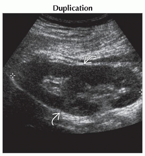
Duplication
Crossed Fused Ectopia
Compensatory Hypertrophy
Multicystic Dysplastic Kidney (MCDK)
Pyelonephritis
Wilms Tumor
Less Common
Multilocular Cystic Nephroma
Mesoblastic Nephroma
Renal Vein Thrombosis, Acute Phase
Trauma
Rare but Important
Infarction
Venolymphatic Malformations
Renal Medullary Carcinoma
Unusual Renal Tumors
ESSENTIAL INFORMATION
Key Differential Diagnosis Issues
Comparison to contralateral kidney is key
Look up renal length when side of abnormality is not apparent
Assess
Renal contour
Corticomedullary differentiation
Contrast enhancement pattern
Presence of cysts or masses
Vascular compromise: Abnormal supply, compression, tumor thrombus
Adjacent spaces and nodes
Helpful Clues for Common Diagnoses
Duplication
Look for band of cortex separating upper and lower poles
Look for 2nd renal pelvis and ureter
Crossed Fused Ectopia
Contralateral kidney will be absent
Occasionally lower renal tissue lies in midline, “J” shape
Part of spectrum of renal ascent and rotation abnormalities
Lower renal moiety ureter will cross to contralateral trigone
Compensatory Hypertrophy
Seen most commonly with contralateral aplasia or MCDK
Also seen following early surgical removal of kidney
Less common with hypoplasia/dysplasia
Multicystic Dysplastic Kidney (MCDK)
Classic type: Conglomerate cysts without discernible renal pelvis
Hydronephrotic type: Central cyst thought to be remnant of obstructed pelvis
High incidence of contralateral renal abnormalities
Ureteropelvic junction obstruction
Vesicoureteral reflux
MCDK initially large, but vast majority shrink over course of years
1/2 of all MCDK have involuted by age 5
12 reported cases of Wilms and renal cell carcinoma occurring in MCDK
National MCDK Registry tracks incidence and behavior
Imaging: Ultrasound and nuclear scan to confirm nonfunction
Pyelonephritis
May have normal echotexture on ultrasound
Look for
Altered corticomedullary interface
Focal hypoechoic area
Decreased perfusion on Doppler exam
Bulge in cortex from focal swelling
Striated nephrogram on CT, MR, or IVP
Poorly enhancing areas on contrast studies
Wedge-shaped photopenic area on DMSA
Imaging
DMSA most sensitive exam
CT & MR next most sensitive
Ultrasound least sensitive but does exclude complications of abscess, perinephric collection, obstruction
Wilms Tumor
Malignant tumor of primitive metanephric blastema
Most common renal tumor in children
Peak incidence ages 2-5
Typically heterogeneous soft tissue
Vascular extension and tumor thrombus common
5-10% are bilateral, associated with nephroblastomatosis
Large tumors grow into perinephric space, periaortic nodes
Metastasize to lung
Cure rate is 90% or better with chemotherapy
Imaging: Ultrasound, CT, MR, nucs (per treatment protocol)
Helpful Clues for Less Common Diagnoses
Multilocular Cystic Nephroma
a.k.a. cystic nephroma
Rare, benign tumor
Bimodal age distribution
Childhood: M:F = 2:1, 3 months to 2 years
Adulthood: M:F = 1:8, 30 years or older
Can mimic cystic Wilms tumor; always removed
Imaging: Ultrasound, CT, MR, nucs (per treatment protocol)
Mesoblastic Nephroma
Most common renal tumor in neonate
Peak age 3 months
Typically solid but can also be cystic
Predominantly benign but can be locally invasive or recur
Imaging: Ultrasound, CT, MR, nucs (per treatment protocol)
Renal Vein Thrombosis, Acute Phase
Obstruction to venous outflow causes renal engorgement, ischemia, and eventually infarction if not relieved
Chronically, kidney scars and atrophies
Imaging: Ultrasound with Doppler, CT angiography, conventional angiography (rarely)
Trauma
Contusion of kidney with global swelling
Perinephric hematomas, urinomas, subcapsular hematomas, etc. can all mimic enlarged kidney
Imaging: CT best in acute setting, ultrasound to follow-up
Helpful Clues for Rare Diagnoses
Infarction
Arterial compromise, particularly from emboli, can cause renal enlargement
Venolymphatic Malformations
Rarely retroperitoneal vascular malformation may involve kidney
Renal Medullary Carcinoma
Rare tumor
Young patients with sickle cell trait
Highly aggressive tumor
Central, infiltrating tumor with caliectasis and regional adenopathy
Metastases often seen at presentation
Poor prognosis
Unusual Renal Tumors
Clear cell carcinoma
Rhabdoid tumor
Primitive neuroectoderm tumor (PNET)
Renal cell carcinoma
Image Gallery
 Longitudinal ultrasound shows a band of parenchyma
 crossing the central sinus fat and dividing the kidney into upper and lower moieties in this 14 year old with a 15 cm long right kidney. crossing the central sinus fat and dividing the kidney into upper and lower moieties in this 14 year old with a 15 cm long right kidney.Stay updated, free articles. Join our Telegram channel
Full access? Get Clinical Tree
 Get Clinical Tree app for offline access

|