Unilateral Hydronephrosis
Sara M. O’Hara, MD, FAAP
DIFFERENTIAL DIAGNOSIS
Common
Ureteropelvic Junction (UPJ) Obstruction
Ureterovesical Junction (UVJ) Obstruction
Ureterocele
Posterior Urethral Valves
Urolithiasis (Stones)
Vesicoureteral Reflux
Less Common
Megaureter
Bladder Mass
Iatrogenic
VUR Post Ureterocele Incision
Deflux Complications
Ureteral Re-Implant Complications
Stent Misplacement or Blockage
Ureteral Ligation During Pelvic Surgery
Rare but Important
Ureteral Fibroepithelial Polyp
ESSENTIAL INFORMATION
Key Differential Diagnosis Issues
Many cases are found prenatally and precisely diagnosed in newborn period
Determining extent or level of dilation is key to narrowing differential
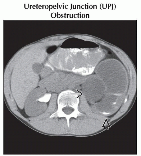
Dilated calyces and disproportionately enlarged renal pelvis often indicate UPJ obstruction
Dilation from top to bottom points to problems at UVJ
Intrinsic UVJ obstruction
UVJ stone
Ureterocele
Megaureter
Bladder mass
Compression from other pelvic mass
Mid-ureteral transitions to normal caliber are unusual; if present, consider
Urolithiasis
Aberrant anatomy: Crossing vessel
Stricture: Postsurgical or radiation related
Compression by adjacent mass
Ureteral mass or polyp is very uncommon
Helpful Clues for Common Diagnoses
Ureteropelvic Junction (UPJ) Obstruction
Most common cause of hydronephrosis in children
Focal narrowing at junction of renal pelvis and ureter
Degree of UPJ obstruction
Varies from mild to severely obstructed
May change with age and hydration state
Obstruction may be intrinsic or extrinsic
Intrinsic likely starts in fetal life
Fibrosis, abnormal innervation, failure of canalization are all suspected etiologies
By the time narrowing is resected, pathologically cannot determine cause
Extrinsic causes are typically crossing vessels, adenopathy, or mass
Imaging of UPJ obstruction
Ultrasound and diuretic renography are mainstays for degree of obstruction, scarring, renal growth, superimposed infection
Treatment of UPJ obstruction
Dismembered pyeloplasty
Ureterovesical Junction (UVJ) Obstruction
2nd most common cause of hydronephrosis in children
Megaureter is specific subtype of UVJ obstruction
Caused by distal adynamic segment
Other causes of UVJ obstruction
Stricture, fibrosis, aberrant anatomy
Compressed distal ureter: Bladder wall hypertrophy, mass, adenopathy, etc.
Imaging: Diuretic renogram, ultrasound, RUG, VCUG, MR urogram
Ureterocele
Congenital cystic dilation of distal submucosal ureter
Location: Intra- or extravesical
Insertion site
Orthotopic at trigone (“simple”)
Ectopic insertion anywhere else, typically medial and distal to trigone
Kidney being drained
Single system; typically simple, intravesicle variety
Duplex system; typically ectopic &/or extravesical
Imaging: VCUG, US, MR urography
Posterior Urethral Valves
Congenital condition
Seen only in boys
Persistent tissue just distal to verumontanum partially obstructs urethra
Unilateral VUR or urinoma protective to contralateral kidney
Better long-term prognosis
Degree of obstruction varies
Severe obstruction seen in fetus and newborn
Secondary oligohydramnios, respiratory and renal insufficiency
Mild obstruction may go undetected for several years
Can present late with renal failure, bladder dysfunction
Imaging: VCUG
Shows valve tissue &/or urethral caliber change
Treatment: Endoscopic valve ablation
Urolithiasis (Stones)
Much less common problem in pediatrics than in adults
Stone types in decreasing frequency
Calcium phosphate or oxalate
Struvite
Uric acid
Cystine
Mixed
Degree of obstruction varies with stone size and location
Imaging: Ultrasound, CT, IVP (rare)
Treatment: Hydration, diuretics, endoscopic basketing, lithotripsy
Vesicoureteral Reflux
Retrograde flow of urine from bladder toward kidneys
Graded from 1 (mild) to 5 (severe)
80% of children outgrow reflux by puberty
Associated infection and renal scarring
Imaging: VCUG, nuclear cystogram, sonocystogram (where ultrasound contrast is available)
Helpful Clues for Less Common Diagnoses
Megaureter
Focal concentric narrowing of extravesical distal ureter 1-3 cm in length
Unknown etiology; theorized causes include
Paucity of ganglion cells
Hypoplasia/atrophy of muscle fibers in distal ureteral segment
Refluxing and nonrefluxing varieties
Imaging: Diuretic renogram, ultrasound, VCUG, MR urography
Treatment: Resection of narrowed segment and re-implantation
Bladder Mass
Rhabdomyosarcoma most common
Inflammatory pseudotumor
Neuroblastoma and transitional cell rare
Imaging: Ultrasound, VCUG, CT, or MR for local extent
Treatment: Resection and ureteral re-implant or diversion
Iatrogenic
Consider whenever there has been recent surgery or invasive procedure
Helpful Clues for Rare Diagnoses
Ureteral Fibroepithelial Polyp
Benign, rare tumor of urothelium
Image Gallery
 Axial CECT after 10-minute delay shows marked dilation of the left renal pelvis
 and calyces, with minimal contrast layering in the calyces and calyces, with minimal contrast layering in the calyces  . Excreted contrast is seen in the right renal pelvis with none in the left. . Excreted contrast is seen in the right renal pelvis with none in the left.Stay updated, free articles. Join our Telegram channel
Full access? Get Clinical Tree
 Get Clinical Tree app for offline access

|