and Marcelo Zugaib4
(1)
São Paulo University, Bauru, Brazil
(2)
Parisian University, Bauru, France
(3)
Member of International Fetal Medicine and Surgery Society, Bauru, Brazil
(4)
Obstetrics, University of São Paulo, Bauru, Brazil
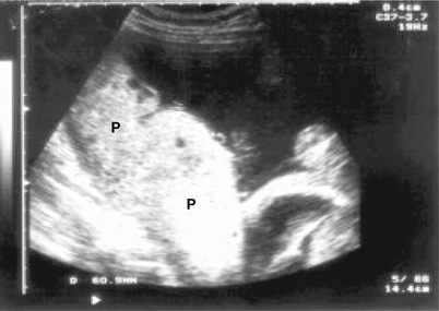
Fig. 15.1
Ultrasound image of a placenta (P) with a thickness of 50 mm (thickened), showing placental involvement in a case of congenital toxoplasmosis

Fig. 15.2
Fetal ascites seen in cross-section of the fetal abdomen at the level of the liver (A). Case of toxoplasmosis confirmed

Fig. 15.3
Cross-section of the cephalic pole showing ventriculomegaly, lateral ventricle (LV) in a case of fetal cytomegalovirus infection
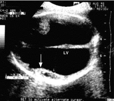
Fig. 15.4
Cross-section of the cephalic pole showing dilatation of the lateral ventricles (LV) and residual brain parenchyma (arrow) in a fetus with toxoplasmosis
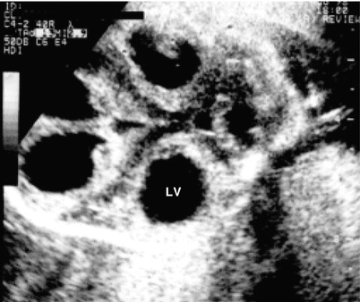
Fig. 15.5
Dilatation of the lateral ventricles (LV) observed in a cross-section of the cephalic pole at the level of the thalamus, in a fetus with toxoplasmosis

Fig. 15.6
Image of the central nervous system of a fetus carrying congenital toxoplasmosis with major dilation of the lateral ventricles, an anechoic image (arrow), and absence of brain parenchyma, which is called hydranencephaly

Fig. 15.7
CT scan of the central nervous system (CNS) of a newborn congenital toxoplasmosis carrier with dilation of the brain ventricles. Anechoic image (arrow)

Fig. 15.8
Fetus with congenital toxoplasmosis demonstrating periventricular calcifications, hyperechoic images (black arrows) on the wall of the lateral ventricle (LV), above the thalamus (white arrow)
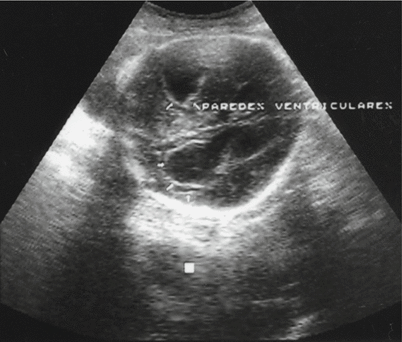
Fig. 15.9
Cross-section of the fetal cephalic pole with periventricular calcifications (arrows) in the context of a fetal infection of cytomegalovirus, noting the presence of lateral ventricle dilation

Fig. 15.10
Part of the central nervous system showing dilatation of the lateral ventricles in a case of fetal toxoplasmosis diagnosed by amniotic fluid analysis through polymer chain reaction. Fetal death at 26 weeks of amenorrhea

Fig. 15.11
Spleen: hypoechoic image localized to the left and behind the stomach, in a cross-section of the fetal abdomen at this level (arrow), which corresponds to splenomegaly in a fetus with positive polymer chain reaction for Toxoplasma in an amniotic fluid sample. Fetal splenomegaly
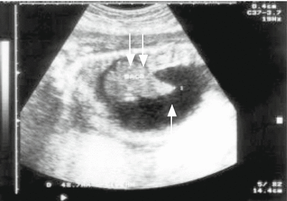
Fig. 15.12
Evaluation of the fetal abdomen at the level of the spleen, showing ascites (single arrow) and assessing the size of the spleen (double arrows)
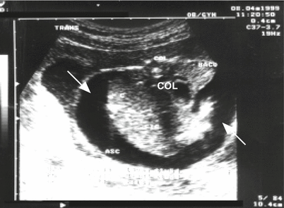
Fig. 15.13
Congenital toxoplasmosis coursing with fetal hydrops. Cross-section of the fetal abdomen at the level of the liver with an anechoic image and ascites (arrow). COL column, ASC ascites
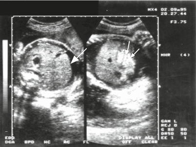
Fig. 15.14
Evaluation of ascites in a cross section of the fetal abdomen at the level of the liver. Anechoic image laminate surrounding the liver (larger arrow). In the fetal abdomen, a solid hyperechoic image, a hyperechoic gut (lower arrows)
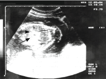
Fig. 15.15
Hyperechoic image observation corresponding to an intestine (black arrows) in a longitudinal cross-section of the fetal abdomen at the level of the liver, in a case of suspected cytomegalovirus


