and Marcelo Zugaib4
(1)
São Paulo University, Bauru, Brazil
(2)
Parisian University, Bauru, France
(3)
Member of International Fetal Medicine and Surgery Society, Bauru, Brazil
(4)
Obstetrics, University of São Paulo, Bauru, Brazil
Neural tube defects (NTDs) belong to a group of fetal malformations with high relevance in perinatal care, because of their prevalence and the possible consequences to the affected individuals. According to the geographic region considered, their incidence varies between 1/1000 and 8/1000 live births, and their recurrence is 2–3%. In Brazil and Latin America, the incidence of NTDs is about 1.2/1000 live births.


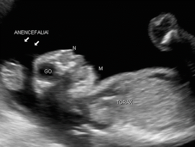


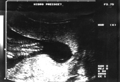


Fig. 4.1
(a, b) Sagittal images of fetal dorse back showing skin (arrows), ossification centers of the posterior vertebral arches (A), and intact vertebral bodies (C)
Fig. 4.2
Sagittal images of the fetal back at a lumbosacral spine level, demonstrating horsetail-like (in Latin, cauda equina) and conus medullaris with normal aspects
Fig. 4.3
Axial image of the normal fetal spine. P = skin, C = vertebral bodies, A = ossification center of the posterior vertebral arch
Fig. 4.4
Coronal plane in the fetal spine revealing the usual proximity of the ossification centers of the lateral vertebral arches (arrows). To the left, the lumbar and sacral bones. To the right, thoracic and cervical spines
Fig. 4.5
Sagittal view of fetus at 11 weeks gestational (CCN = CRL) showing exposition of brain parenchyma (arrows exencephaly) without coating of skull cap (acrania) such conditions are observed in the early stages of anencephaly
Fig. 4.6
Sagittal view of fetus at 12 weeks of gestational age showing exposition of brain parenchyma (arrows exencephaly) without coating of skull cap (acrania) such conditions are observed in the early stages of anencephaly
Fig. 4.7
Axial (left) and sagittal (right) images of the fetal cephalic pole with acrania and exencephaly (arrows). T = thorax, F = face
Fig. 4.8
Sagittal image of the fetal cephalic pole at 16 weeks’ gestational age, showing, during this phase, the aspect of acrania with exencephaly (arrows). C = spine, T = thorax, F = face
Fig. 4.9
Axial image of the fetal cephalic pole at 16 weeks of gestational age, showing, during this phase, the aspect of acrania with exencephaly (arrows). C = spine, T = thorax, F = face
Fig. 4.10
Sagittal image of the fetal cephalic pole revealing the absence of the coating of the skullcap (arrows) with protruding eye globes (GO), a typical aspect observed in anencephaly cases
Fig. 4.11
Coronal slice, tangential to the face of an anencephalic fetus (GL = eyeball) showing a batrachian aspect. BOCA = mouth
Fig. 4.12
Sagittal section showing the abrupt termination of bone structures in the fetal head and no brain parenchyma (anencephaly, arrows) in an anencephalic fetus. J = fetal knee, TO = thorax, N = nostrils, M = chin
Fig. 4.13
Coronal section identifying anencephaly (right arrows) associated with acrania and spina bifida (left) featuring cervical craniorachischisis (arrows) in pregnancy with gestational age of 12 weeks and 4 days. O = eyeballs, m = chin
Fig. 4.14
Coronal section of the lumbar spine of a fetus at 14 weeks of gestational age, demonstrating removal of the ossification centers of the posterior vertebral arches (arrows) in a case of open spina bifida
Fig. 4.15
Axial section of the lumbar spine of a fetus at 14 weeks’ gestational age, demonstrating removal of the ossification centers of the posterior vertebral arches (arrows) in a case of open spina bifida
Fig. 4.16
Axial section of the fetal cephalic pole showing the appearance of early hydrocephalus in the context of open spina bifida. Note the choroid plexus (CP) hanging loose, away of the ventricular walls (arrow) very close to the cranial bone plate, with thinning of the brain parenchyma
Fig. 4.17
Coronal section of fetal back bones demonstrating the open spina bifida in lumbosacral region (arrows)


