Sagittal section of a fetus showing measurement of crown–rump length.
Algorithm for a structural survey in early pregnancy
Brain
Cranial bone ossification and the integrity of the skull should be noted to exclude severe anomalies like acrania. The hemispheres should appear symmetrical and separated by a clear midline falx cerebri and interhemispheric fissure. By 7 weeks, a sonolucent area can be seen in the cephalic pole. By 9 weeks, a convoluted pattern of three primary cerebral vesicles is noted, followed by the appearance of brightly echogenic choroid plexus filling the lateral ventricles by 11 weeks. It is difficult to assess the integrity of the cerebellum, cavum septum pellucidum and corpus callosum at this gestation (Figures 5.2 and 5.3).
A BPD measurement less than the 10th centile may be a subtle sign of open spina bifida[7].
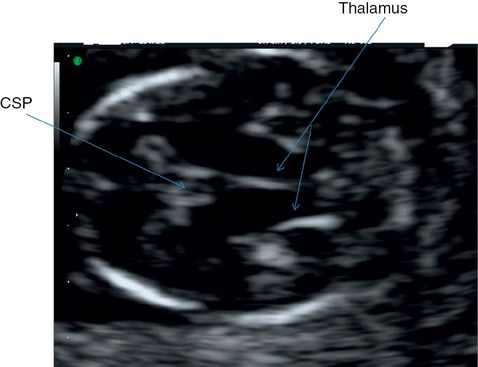
Transverse section through fetal head showing midline echo, thalamus and cavum septum pellucidi (CSP).
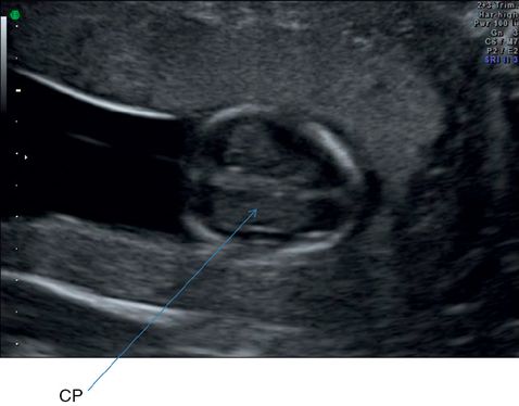
Transverse section through brain ventricles demonstrating relatively prominent-appearing choroid plexus (CP).
Neck
One of the biggest advances in prenatal diagnosis is the measurement of nuchal translucency (NT). The correct NT should be obtained by adopting and ensuring the following criteria are met.
Obtain a sagittal section of the fetus and magnify the image in order to include only the fetal head and upper thorax. The true sagittal section will demonstrate an echo from the tip of the nose, a second echo from the nasal bone and a square and well-defined echo from the maxilla.
Fetal spine and neck should be in a neutral position with a pool of amniotic fluid between the chin and the upper chest. Hyperflexion or hyperextension will lead to erroneous measurement.
Calipers should be placed correctly, with the horizontal portion of the + marker exactly on the inner borders of the lines demarcating the nuchal space or NT. It should be placed perpendicular to the long axis of the fetus at the widest portion of the NT space. The amnion should be seperately identified to avoid mistaking it for the fetal skin (Figure 5.4).
If more than one measurement meeting all the criteria is obtained, the maximum one should be recorded and used for aneuploidy risk assessment.
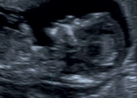
A midline sagittal section showing measurement of nuchal translucency.
Face
An attempt should be made to look at the face in coronal plane. If clear views are not obtained, it should be re-assessed at the second-trimester scan[1].
Heart
A four-chamber view of the heart can be obtained starting from 10 to 13 weeks[8]. Complete examination of the fetal heart in the first trimester can be challenging due to the smaller size. As a minimum, the position of the heart and the four-chamber view should be noted. In case of any suspected abnormality, a detailed scan is warranted in the second trimester for further evaluation.
Abdomen
The physiologic umbilical hernia is present up to 11 weeks, and should be differentiated from gastroschisis and omphalocoele. Kidneys and bladder should be noted. Kidneys appear in the paraspinal region as bean-shaped structures. The bladder can be visualized in the normal site as a hypoechoic area (Figures 5.5–5.8).
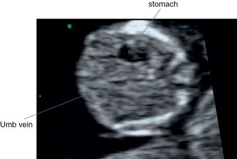
Transverse section through fetal abdomen shows the stomach and an oblique section of the umbilical vein. Longitudinal section of a single rib on either side and a cross-section of the spine is also visible.
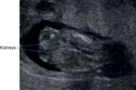
Coronal section through fetal trunk showing a section of the spine and longitudinal section of the fetal kidneys.
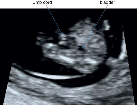
Transverse section through fetal lower abdomen showing umbilical cord insertion and the bladder.
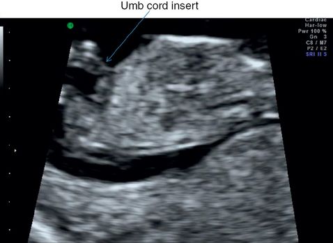
Longitudinal section of fetal trunk showing umbilical cord insertion and the anterior abdominal wall.
Spine
The role of the first-trimester scan in detecting small defects of the spine is limited; however, severe cases of spinal abnormalities may be detected by a systematic examination[9]. Careful observation should be undertaken to visualize an intact skin covering over the spine (Figure 5.9).
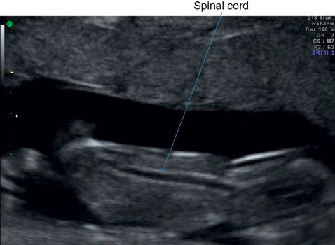
Longitudinal section through fetal spine to show skin covering. The spinal cord is visible as a tubular sonolucent structure. Faint echogenic vertebrae are also visible.
Limbs
Note the presence of four limbs with three bony segments in each of the upper and lower limbs with normal orientation of the two hands and feet (Figure 5.10).
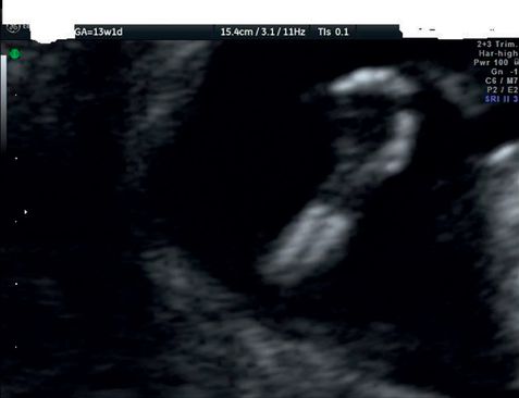
Longitudinal section through fetal forearm and hand showing both long bones and five digits.
Checklist for examination of fetal anatomy in the first trimester[1]
| Brain | Integrity of skull Midline falx Choroid plexus – filled Ventricles |
| Face | Eyes with lens Nasal bone Normal profile/mandible Intact lips |
| Neck | Nuchal translucency thickness Any fluid-filled collections |
| Spine | Longitudinal and axial view of vertebrae Skin integrity |
| Chest | Symmetrical lung fields No effusions or masses Regular cardiac activity Four-chamber view Check situs visceralis with stomach and heart on the left side of the abdomen Diaphragm |
| Abdomen | Stomach in left upper quadrant Intact abdominal wall Normal cord insertion after 12 weeks Kidney and bladder |
| Extremities | Four limbs each with three segments Hands and feet with normal orientation Femoral length |
| Placenta and amniotic fluid volume |
Limitations
One of the limitations of the first-trimester anomaly scan as a routine screening method in a low-risk population is the poor sensitivity for certain anomalies like those of the heart, spine and brain due to the stages of embryologic development. Therefore, even if an early fetal anomaly scan were offered, the second-trimester scan should not be abandoned. Secondly, in women with raised body mass index, the image quality in the transabdominal scan may be compromised. A transvaginal approach will improve the sensitivity, but the patient’s acceptance may be a limiting factor.
The routine second-trimester scan
The second-trimester anomaly scan is offered between 18+0 weeks to 20 weeks and 6 days of gestation in most countries. The timing is appropriate for detailed evaluation of most of the anatomical structures. Although not the ideal gestation for dating the pregnancy, it can be useful in dating in the case of a missed opportunity for a first-trimester scan.
Diagnostic value of routine ultrasound scan in the second trimester
(a) Lethal anomalies[10]
| Anencephaly | 97.6% |
| Trisomy 18 | 68.4% |
| Trisomy 13 | 50.0% |
| Hypoplastic left heart | 54.5% |
| Bilateral renal agenesis | 90.0% |
| Lethal musculoskeletal disorders | 33.3% |
(b) Possible survival and long-term morbidity[10]
| Spina bifida | 66.3% |
| Hydrocephalus | 68.9% |
| Encephalocele | 90.9% |
| Holoprosencephaly | 72.7% |
| Down’s syndrome | 14.6% |
| Complex cardiac malformations | 21.3% |
| Atrioventricular septal defect | 12.9% |
| Nonlethal dwarfism | 100% |
| Anterior abdominal wall defect | 89.5% |
| Gastroschisis | 94.1% |
| Exomphalos | 84.6% |
| Congenital diaphragmatic hernia | 47.9% |
| Tracheoesophageal atresia | 7.4% |
| Small bowel obstruction/atresia | 40.6% |
| Congenital cystic adenomatoid malformation | 100% |
| Renal dysplasia (bilateral) | 84.2% |
| Multiple abnormality/syndrome | 77.8% |
| Obstructive uropathy | 100% |
| Pleural effusion or hydrothorax | 100% |
(d) Anomalies associated with possible short term/immediate morbidity[10]
| Facial clefts | 13.8% |
| Talipes | 20.1% |
| Atrial/ventricular septal defect | 6.3% |
| Isolated valve anomalies | 22.7% |
| Renal dysplasia (unilateral) | 86.7% |
A stepwise approach for a routine second-trimester anomaly scan is listed below.
(1) Confirm viability.
(2) Order of pregnancy.
(3) Placentation.
(4) Fetal biometry.
(5) Systematic structural survey.
(6) Amniotic fluid assessment.
Fetal biometry
For assessment of the fetal size and growth, the standard measurements used are the HC, BPD, abdominal circumference and femoral length.
Head circumference and biparietal diameter
Both the HC and BPD measurements can be obtained from the same plane; the transventricular plane or the transthalamic plane can be used for obtaining these measurements. The HC can be measured by using the ellipse method by placing the ellipse around the outside of the skull bone echoes. HC measurement is more reliable than the BPD in cases of abnormal shape of the head.
The BPD can be measured by two techniques – the outer-edge to inner-edge technique or the outer-edge to outer-edge by placing the calipers at the widest part of the skull. It is important to ensure that the placement of the calipers corresponds to the technique described on the reference chart, especially for the BPD (outer-to-outer or outer-to-inner) (Figures 5.11 and 5.12).
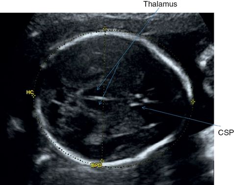
Transthalamic section of fetal head showing measurement of head circumference (HC) and biparietal diameter (BPD).
A cross-sectional view of the head is obtained at the level of thalami with a short midline echo equidistant from the proximal and distal skull echoes. The structures that should be identified in the midline anteriorly to posteriorly are the cavum septum pellucidum (CSP), the thalami and basal cisterns[12].
Stay updated, free articles. Join our Telegram channel

Full access? Get Clinical Tree


