Tumors of the Digestive Tract

Chapter 337 Tumors of the Digestive Tract


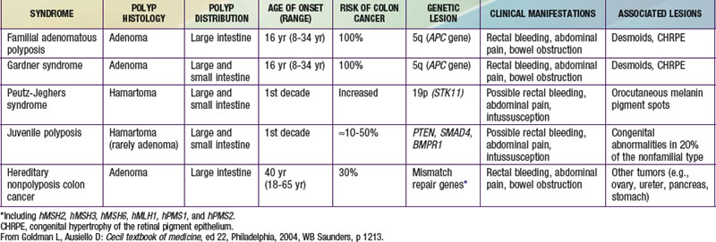
Tumors of the digestive tract are mostly polypoid. They are also commonly syndromic tumors and tumors with known genetic identification (see imageTable 337-1 on the Nelson Textbook of Pediatrics website at www.expertconsult.com). They usually manifest as painless rectal bleeding, but they can serve as lead points for intussusception.


Hamartomatous Tumors


Hamartomas are benign tumors composed of tissues that are normally found in an organ but that are not organized normally. Juvenile, retention, or inflammatory polyps are hamartomatous polyps, which represent the most common intestinal tumors of childhood, occurring in 1-2% of children. Patients generally present in the 1st decade, most often at age 2-5 yr, and rarely at <1 year. Polyps may be found anywhere in the gastrointestinal (GI) tract, most commonly in the colon or rectum; they are often solitary but may be multiple.


Histologically, juvenile polyps are composed of hamartomatous collections of mucus-filled glandular and stromal elements with inflammatory infiltrate, covered with a thin layer of epithelium. These polyps are often bulky, vascular, and prone to bleed as their growth exceeds their blood supply with resultant mucosal ulceration, or autoamputation with bleeding from a residual central artery.


Patients often present with painless rectal bleeding after defecation. Bleeding is generally scant and intermittent; rarely iron deficiency anemia is the chief presenting symptom. Extensive bleeding can occur but is generally self-limited, requiring supportive care until the bleeding stops spontaneously after autoamputation. Occasionally endoscopic polypectomy is required for control of bleeding. Abdominal pain or cramps are uncommon unless associated with intussusception. Patients can present with prolapse, with a dark, edematous, pedunculated mass protruding from the rectum. Mucus discharge and pruritus are associated with prolapse.


Patients presenting with rectal bleeding require thorough work-up; differential diagnosis includes anal fissure, other intestinal polyposis syndromes, Meckel’s diverticulum, inflammatory bowel disease, intestinal infections, Henoch-Schönlein purpura, or coagulopathy.


Diagnosis and therapy are best accomplished via endoscopy. Polyps may be visualized via air-contrast barium enema, but this provides no therapeutic advantage and is uncomfortable and usually performed without sedation or anesthesia. Colonoscopy affords opportunity for biopsy, polypectomy by snare cautery, and visualization of synchronous lesions; up to 50% of children have ≥1 additional polyp, and ∼20% may have >5 polyps. Retrieved polyps should be sent for histologic evaluation for definitive diagnosis.


Peutz-Jeghers Syndrome


Peutz-Jeghers syndrome (PJS) is a rare autosomal dominant disorder (incidence, ∼1 : 120,000) characterized by mucocutaneous pigmentation and extensive GI hamartomatous polyposis. Macular pigmented lesions may be dark brown to dark blue and are found primarily around the lips and oral mucosa, although these lesions may also be found on the hands, feet, or perineum (Fig. 337-1). Lesions can fade by puberty or adulthood.


Polyps are primarily found in the small intestine (in order of prevalence: jejunum, ileum, duodenum) but may also be colonic or gastric. Histologically polyps are defined by normal epithelium surrounding bundles of smooth muscle arranged in a branching or frondlike pattern. Symptoms arising from GI polyps in PJS are similar to those of other polyposis syndromes, namely bleeding and abdominal cramping from obstruction or recurrent intussusception. Patients can require repeated laparotomies and intestinal resections.


The diagnosis of PJS is made clinically in patients with histologically proven hamartomatous polyps if two of three conditions are met: positive family history with an autosomal dominant inheritance pattern, mucocutaneous hyperpigmentation, and small bowel polyposis. Genetic testing can reveal mutations in STK11 (LKB1; 19p13.3), a serine-threonine kinase that acts as a tumor-suppressor gene. Up to 94% of patients with clinical characteristics of PJS have a mutation at this locus. Only 50% of patients with PJS have an affected family member, suggesting a high rate of spontaneous mutations.

Stay updated, free articles. Join our Telegram channel

Jun 18, 2016 | Posted by in PEDIATRICS | Comments Off on Tumors of the Digestive Tract

Full access? Get Clinical Tree

Get Clinical Tree app for offline access