Fig. 9.1
Position of the patient (P), surgeon (S), camera holder (C), audiovisual equipment (AV), scrub nurse (N), and anesthetist (A) for transposition of right renal vessels
Port Position
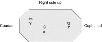
One primary umbilical port and two working ports are required. The port positions are shown in Fig. 9.2. A trick to facilitate umbilical primary port access by the open technique is to tilt the table away from the surgeon to make the patient more supine. Once access is obtained, the table can be returned to its original position and the patient in the renal position.


Fig. 9.2
Port position X (primary) and Y–Z (secondary ports)
Operative Technique
The ascending or descending colon is reflected medially to expose the perirenal fascia and quite often the bulging renal pelvis.
The perirenal fascia is incised and reflected medially and the adventitia over the pelvis cleared.
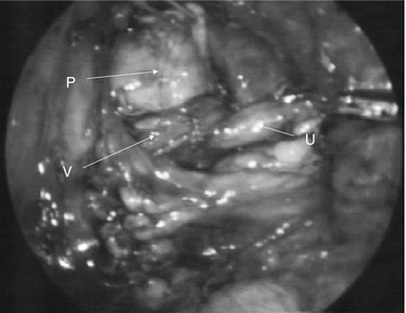
The pelvis is traced inferiorly to the PUJ, or the ureter is traced superiorly to expose the lower pole vessels (Fig. 9.3).

Fig. 9.3
Renal pelvis (P), ureter (U), and vessels (V) exposed (Reprinted from Godbole et al. [3]. Copyright© 2006, with permission from Elsevier)
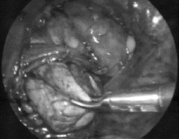
If lower pole vessels are identified, with a combination of scissors/bipolar hook, the pelvis and ureter are fully mobilized so that they are completely free from the lower pole vessels (Fig. 9.4). This can be demonstrated by the “shoe shine” maneuver as seen in the accompanying video.

Fig. 9.4
Renal pelvis (P) fully mobilized (Reprinted from Godbole et al. [3]. Copyright© 2006, with permission from Elsevier)
Stay updated, free articles. Join our Telegram channel

Full access? Get Clinical Tree


