Transmissible Spongiform Encephalopathies

Chapter 270 Transmissible Spongiform Encephalopathies


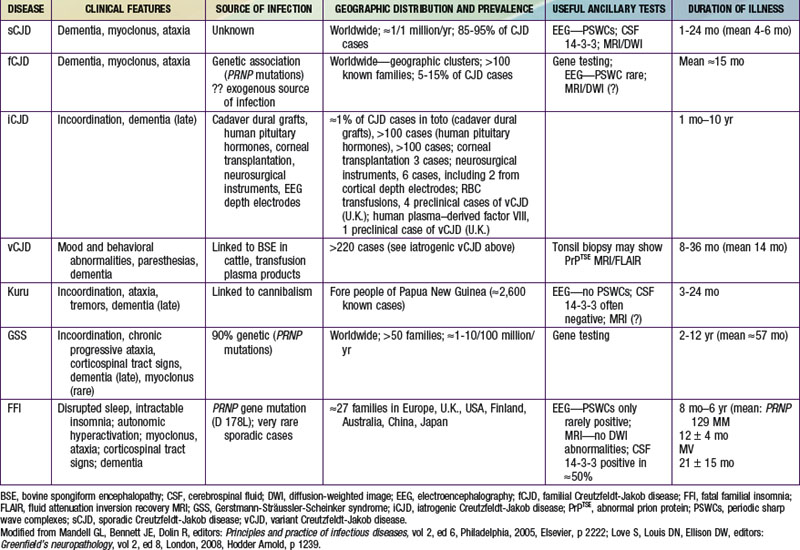
The transmissible spongiform encephalopathies (TSEs) are slow infections of the human nervous system, consisting of at least 4 diseases of humans (see Table 270-1 imageon the Nelson Textbook of Pediatrics website at www.expertconsult.com): kuru; Creutzfeldt-Jakob disease (CJD) with its variants—sporadic CJD (sCJD), familial CJD (fCJD), iatrogenic CJD (iCJD), and new-variant or variant CJD (vCJD); Gerstmann-Sträussler-Scheinker syndrome (GSS); and fatal familial insomnia (FFI), or the even more rare sporadic fatal insomnia syndrome. TSEs also affect animals; the most common and best-known TSEs of animals are scrapie in sheep, bovine spongiform encephalopathy (BSE or mad cow disease) in cattle, and a chronic wasting disease (CWD) of deer, elk, and moose found in parts of the USA and Canada. All TSEs have similar clinical manifestations and histopathology, and all are “slow” infections with very long asymptomatic incubation periods (often years), durations of several months or more, and overt disease affecting only the nervous system. The most striking neuropathologic change that occurs in each TSE, to a greater or lesser extent, is spongy degeneration of the cerebral cortical gray matter.



Etiology


The TSEs are transmissible to susceptible animals by inoculation of tissues from affected subjects. Although the infectious agents replicate in some cell cultures, they do not achieve the high titers of infectivity found in brain tissues or cause recognizable cytopathic effects in cultures. Most studies of TSE agents have used in vivo assays, relying on the transmission of typical neurologic disease to animals as evidence that the agent was present and intact. Inoculation of susceptible recipient animals with small amounts of infectious TSE agent results, months later, in the accumulation in tissues of large amounts of agent with the same physical and biologic properties as the original agent. The TSE agents display a spectrum of extreme resistance to inactivation by a variety of chemical and physical treatments that is unknown among conventional pathogens. This characteristic, as well as their partial sensitivity to protein-disrupting treatments and their consistent association with abnormal isoforms of a normal host-encoded protein (prion protein or PrP), stimulated the hypothesis that the TSE agents are probably subviral in size, composed of protein, and devoid of nucleic acid.


The term prion (for proteinaceous infectious agent) has been suggested as an appropriate name for such agents. The prion hypothesis proposes that the molecular mechanism by which the pathogen-specific information of TSE agents is propagated involves a self-replicating change in the folding host-encoded PrP associated with a transition from an α-helix–rich structure in the native protease-sensitive conformation (cellular PrP or PrPC) to a β-sheet–rich structure in the protease-resistant conformation associated with infectivity. The existence of a 2nd host-encoded protein—termed “protein X”—that participates in the transformation was also postulated to explain certain otherwise puzzling findings.


The prion hypothesis is not universally accepted; it relies on the postulated existence of a genome-like coding mechanism based on differences in protein folding that have not been satisfactorily explained at a molecular level. In addition, it has yet to account convincingly for the many biologic strains of TSE agent that have been observed, although strain-specific differences in the abnormal forms of the PrP have been found and proposed as providing a molecular basis for the coding. It fails to explain why pure PrP uncontaminated with nucleic acid from an infected host has not transmitted a convincingly typical spongiform encephalopathy associated with a serially self-propagating agent. Also troubling, in several experimental models, abnormal PrP and infectivity were not consistently associated. If the TSE agents ultimately prove to consist of protein and only protein, without any obligatory nucleic acid component, then the term prion will indeed be appropriate. If the agents are ultimately found to contain small nucleic acid genomes, then they might better be considered atypical viruses, for which the term virino has been suggested. Until the actual molecular structure of the infectious TSE pathogens and the presence or absence of a nucleic acid genome are rigorously established, it seems less contentious to continue calling them TSE agents, although many authorities use the term prion.


The 1st evidence that abnormal proteins are associated with the TSE was morphologic: scrapie-associated fibrils (SAFs) were found in extracts of tissues from patients and animals with spongiform encephalopathies but not in normal tissues. SAFs resemble but are distinguishable from the amyloid fibrils that accumulate in the brains of patients with Alzheimer disease. A group of antigenically-related protease-resistant proteins (PrPs) proved to be components of SAF and to be present in the amyloid plaques found in the brains of patients and animals with TSEs. The abnormal forms of PrP are variously designated PrPSc (scrapie-type PrP), PrP-res (protease-resistant PrP), PrPTSE (TSE-associated PrP), or PrPD (disease-associated PrP) by different authorities.


It remains unclear whether abnormal PrP constitutes the complete infectious particle of spongiform encephalopathies, is a component of those particles, or is a pathologic host protein not usually separated from the actual infectious entity by currently used techniques. The demonstration that PrP is encoded by a normal host gene seemed to favor the last possibility. Several studies have suggested that agent-specific pathogenic information can be transmitted and replicated by different conformations of a protein with the same primary amino acid sequence in the absence of agent-specific nucleic acids. Properties of 2 fungal proteins were found to be heritable without encoding in nucleic acid, although those properties have not been transmitted to recipient fungi as infectious elements. Whatever its relationship to the actual infectious TSE particles, PrP clearly plays a central role in susceptibility to infection, because the normal PrP must be expressed in mice and cattle if they are to acquire a TSE or to sustain replication of the infectious agents. Furthermore, inherited variations in PrP phenotype are associated with increased susceptibility to vCJD and with occurrence of fCJD.


PrPs are glycoproteins; protease-resistant PrPs have the physical properties of amyloid proteins. The PrPs of several species of animals are very similar in their amino acid sequences and antigenicity but are not identical in structure. The primary structure of PrP is encoded by the host and is not altered by the source of the infectious agent provoking its formation. The function of the ubiquitous protease-sensitive PrP precursor (designated PrPC or PrP-sen, for protease-sensitive PrP) in normal cells is unknown; it binds copper and may play some role in normal synaptic transmission, but it is not required for life or for relatively normal cerebral function in mice and cattle. As noted, expression of PrP is required both for development of scrapie disease and for replication of the transmissible scrapie agent in animals. The degree of homology between amino acid sequences of PrPs in different animal species may correlate with the “species barrier” that affects susceptibility of animals of 1 species to infection with a TSE agent adapted to grow in another species.


Attempts to find particles resembling those of viruses or virus-like agents in brain tissues of humans or animals with spongiform encephalopathies have been unsuccessful. Peculiar tubulovesicular structures reminiscent of some viruses have been seen in thin sections of TSE-infected brain tissues and cultured cells but not in normal cells. It has never been established that those structures are associated with infectivity.


It has been claimed that 2 other human diseases, familial Alzheimer disease of adults and Alpers disease of young children, may be caused by infections with agents similar to those causing the spongiform encephalopathies. The latter is a convulsive disorder associated with hemiatrophy and status spongiosus of the cerebral gray matter. Attempts to confirm these claims by transmission of disease to experimental animals failed.


Epidemiology


Kuru once affected many children ≥4 yr of age, adolescents, and young adults (mainly women) living in 1 limited area of Papua New Guinea. The complete disappearance of kuru among people born after 1957 suggests that the practice of ritual cannibalism (thought to have ended that year) was probably the only mechanism by which the infection was spread in Papua New Guinea.


CJD, the most common human spongiform encephalopathy, was formerly thought to occur only in older adults; however, iCJD and, much more rarely, sCJD have affected adolescents and young adults. GSS and the insomnia syndromes have not been diagnosed in children or adolescents. Variant CJD has a peculiar predilection for younger people; of 174 cases of CJD reported to date in the U.K., all except 23 were in people younger than 40 yr of age and 22 were under 20 yr of age. CJD has been recognized worldwide, at yearly rates of 0.25 to 2 cases/million population (not age-adjusted), with foci of considerably higher incidence among Libyan Jews in Israel, in isolated villages of Slovakia, and in other limited areas. Sporadic CJD has not been convincingly linked to any common exposure, and the source of infection remains unknown. Epidemiologic surveys have investigated several hypothetical mechanisms of spread of CJD. Person-to-person spread has been confirmed only for iatrogenic cases. Spouses and household contacts of patients are at very low risk of acquiring CJD, although 2 instances of conjugal CJD have been reported. However, medical personnel exposed to brains of patients with CJD may be at some increased risk; at least 20 health care workers have been recognized with the disease.


The striking resemblance of CJD to scrapie prompted a concern that infected sheep tissues might be a source of spongiform encephalopathy in humans. No reliable epidemiologic evidence suggests that exposure to potentially scrapie-contaminated animals, meat, meat products, or experimental preparations of the scrapie agent have transmitted a TSE to humans. The potential of the CWD agent to infect human beings has not been demonstrated but remains under investigation; deer, elk, and moose in 16 U.S. states and 2 Canadian provinces have been naturally infected and monkeys have been experimentally infected with the CWD agent. Exposure to contaminated meat, including venison from animals infected with the CWD agent, has not been implicated as a risk factor for sporadic CJD.


The outbreak of BSE among cattle (possibly infected by eating scrapie-agent–contaminated meat-and-bone meal added to feed) was 1st recognized in the United Kingdom in 1986, later reported in native cattle of 24 other countries, including Canada and the USA. The finding of a new TSE in ungulate and feline animals in British zoos and later in domestic cats raised a fear that some TSE agent (probably a strain of the scrapie agent), having crossed the species barrier from sheep to cattle, had acquired a broadened range of susceptible hosts, posing a potential danger for humans. That is the plausible explanation for the occurrence of vCJD, 1st described in adolescents in Britain in 1996 and as of December 2010 affecting at least 174 people in the U.K. (not counting several with evidence of “preclinical” vCJD infection), 25 in France, 5 in Spain, 4 in Ireland, 3 in the Netherlands, 2 each in Italy and Portugal, and single cases in Italy, Japan, and Saudi Arabia. Variant CJD has also occurred in former U.K. residents living in Canada (1 case) and the USA (2 cases); a 3rd case of vCJD in the USA was reported in a former resident of Saudi Arabia, a country that has not recognized BSE but might have imported contaminated meat products.


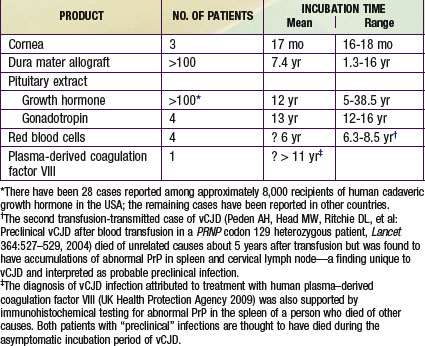
Iatrogenic transmissions of CJD have been recognized for >30 yr (Table 270-2). Such accidental transmissions of CJD have been attributed to use of contaminated neurosurgical instruments or operating facilities, use of cortical electrodes contaminated during epilepsy surgery, injections of human cadaveric pituitary growth hormone and gonadotropin, and transplantation of contaminated corneas and allografts of human dura mater used as a surgical patching material. Pharmaceuticals and tissue grafts derived from or contaminated with human neural tissues, particularly when obtained from unselected donors and large pools of donors, pose special risks.


Stay updated, free articles. Join our Telegram channel

Jun 18, 2016 | Posted by in PEDIATRICS | Comments Off on Transmissible Spongiform Encephalopathies

Full access? Get Clinical Tree

Get Clinical Tree app for offline access