19.2 Thyroid disorders
Normal thyroid function throughout infancy, childhood and adolescence is essential for a normal developmental and physiological outcome. Thyroid disease is one of the most common groups of endocrine disorders in childhood and adolescence, with approximately 1–2% of all children having a thyroid disorder at some time. Therefore, knowledge of thyroid disease and its management is fundamental to paediatric medicine.
Thyroid physiology
The thyroid gland removes iodide from the bloodstream, combines it with tyrosine and releases iodinated tyrosine to the peripheral tissues. The thyroid gland is able to trap iodide and synthesize iodothyronine from 70 days’ gestation. Release of thyroxine, however, does not occur until 18–20 weeks’ gestation. Thyroid gland growth is regulated by thyroid-stimulating hormone (TSH) released from the anterior pituitary gland, which is in turn regulated by thyrotropin-regulating hormone (TRH) released from the hypothalamus. These regulating hormones are in turn controlled by negative feedback from tri-iodothyronine (T3), the active metabolite of the major thyroid hormone thyroxine (or tetra-iodothyronine, T4).
The thyroid gland is extremely effective at trapping serum iodide, with a concentration gradient from thyroid to serum of 30–40-fold. This gradient increases in times of iodide deficiency. Once trapped, iodide is oxidized to iodine and organification occurs. Organification is the iodination of thyroglobulin-bound tyrosyl residues to form mono-iodotyrosine (MIT) and di-iodotyrosine (DIT).
Organification and iodide oxidation (to iodine) are catalysed by thyroid peroxidase. Thyroid peroxidase couples the iodotyrosines to form iodothyronines within the thyroglobulin molecule, resulting in T4 and T3. In the absence of iodine deficiency, the T4 : T3 synthesis ratio is 10–20 : 1. In adults, the release rate of T4 to T3 is 3 : 1. Once released, both hormones bind to thyroxine-binding globulin (TBG). Some 80% of circulating T3 results from de-iodination of T4 in peripheral tissues. Thyroid hormones bind to a nuclear receptor. T3 binds to this receptor with 10 times the affinity of T4. Once bound, thyroid hormones regulate gene transcription, increasing cytoplasmic proteins, which stimulate mitochondrial activity, thus increasing metabolic rate.
Disorders of thyroid function in childhood can be divided into the following categories:
Hypothyroidism
Congenital
Screening for congenital hypothyroidism has been performed in most developed countries for the past 15–20 years. In Australia, screening for congenital hypothyroidism, phenylketonuria and cystic fibrosis occurs on day 3–5 of life. Because of such screening the clinical picture of ‘cretinism’ (the later effects of congenital hypothyroidism, including severe intellectual disability) thankfully is now rarely seen.
Incidence
The incidence of congenital hypothyroidism is 1 in 3000–5000, with some geographical variation.
Aetiology and genetics
Some 75% of cases are due to dysgenesis (agenesis, ectopia), 10% to dyshormonogenesis, 5% to hypothalamic–pituitary deficiency (central hypothyroidism) and 10% to transient hypothyroidism (iodine exposure, maternal antithyroid antibodies, etc.). Although thyroid disease appears to be sporadic in the majority of children born with hypothyroidism, evidence for a genetic component is increasing. Hypothyroidism is familial in up to 2% of cases, and children with congenital hypothyroidism have a higher incidence of associated abnormalities (cardiac, renal, hip dysplasia) than the general population.
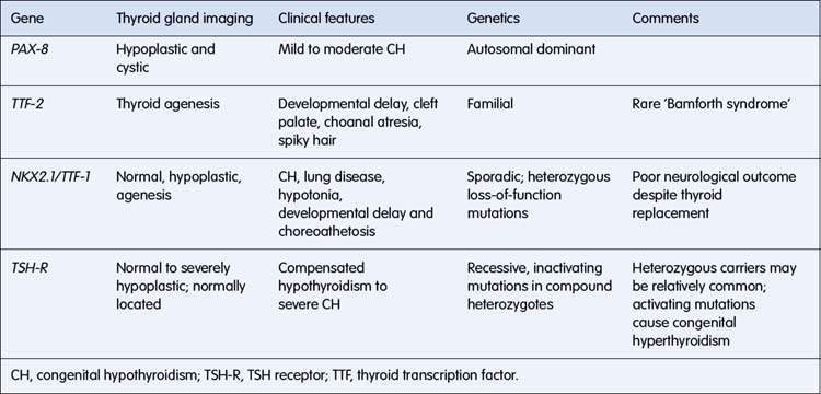
Studies in mouse models with congenital defects of thyroid development have provided the basis for molecular genetic studies in humans with congenital hypothyroidism. Mutations have been described in a number of genes, resulting in absent, misplaced, hypoplastic or unresponsive glands (Table 19.2.1). In some instances, a specific phenotype can be recognized, and prognosis is affected. For example, in individuals with the NKX2.1 mutation, neurological outcome is poor despite early thyroxine treatment.
Table 19.2.1 Mutations in genes involved in thyroid development resulting in congenital hypothyroidism

Hypothyroid patients with normally located and normally sized glands have defects in thyroid hormone biosynthesis. Recessive mutations in thyroglobulin, thyroid peroxidase, pendrin (causing Pendred syndrome – sensorineural deafness and hypothyroidism) and sodium/iodide symporter (NIS) genes have been described. Mutations in the TRH receptor gene, TSH β subunit and transcription factors regulating pituitary development have also been described in some individuals with central hypothyroidism.
Clinical picture
Often the condition is subclinical and is detected on routine screening. Clinical features that should be looked for are jaundice, dry skin, a hoarse cry, puffy face, prominent tongue, listlessness, umbilical hernia, hypothermia, bradycardia and failure to thrive.
Investigation results
An unconjugated hyperbilirubinaemia (due to glucuronyl transferase deficiency) is common. A raised level of TSH detected on testing of a heel-prick drop of blood collected on filter paper on day 3–5 of life is seen in primary hypothyroidism.
Management
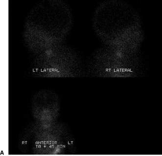
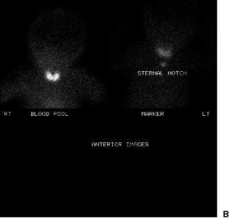
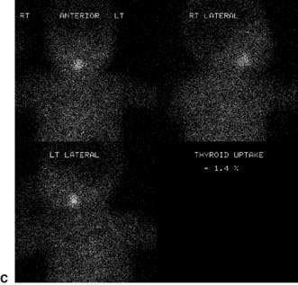
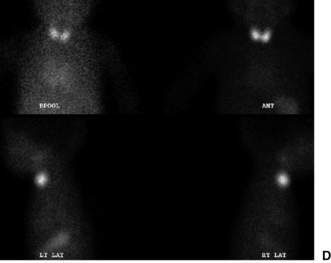
Confirmatory investigations are needed if the screening tests suggest an abnormality: repeat T4 and TSH; thyroid scan (showing absent, lingual or increased uptake of radioisotope), X-ray distal femoral epiphysis (absence implying prolonged/prenatal hypothyroidism), and assessment and imaging of the pituitary gland if indicated. Treatment involves commencement of therapy (thyroxine replacement at 8–10 μg/kg daily). Thyroid imaging results for congenital hypothyroidism of varying causes are shown in Figure 19.2.1.


Fig. 19.2.1 Thyroid uptake scan appearances in congenital hypothyroidism. (A) Thyroid agenesis. No functioning thyroid tissue present in the neck or in the usual ectopic sites. (B) Dyshormonogenesis. The radioangiogram reveals relatively increased thyroid perfusion. The uptake of pertechnetate in 20 min is 14% (normal = 2–5%). The thyroid scan reveals a diffuse goitre normally located in the neck. (C) Lingual thyroid. There is no evidence of perfused thyroid tissue in the neck. The thyroid scan reveals a prominent midline lingual thyroid. The uptake of pertechnetate in 20 min is 1% (normal = 2–5%). (D) Normal thyroid. The radioangiogram of the head, neck and upper torso is unremarkable. The uptake of pertechnetate in 20 min is 5% (normal = 2–5%). The thyroid scan reveals a normally located bilobed gland.
Acquired
Acquired hypothyroidism in the child or adolescent is relatively uncommon. In iodine-sufficient regions of the world the most common cause is autoimmune thyroiditis. Accordingly, acquired hypothyroidism is seen twice as commonly in females as in males, usually manifesting in early puberty.
Prevalence
Between the ages of 1 and 18 years, the prevalence is 1.2%. Acquired hypothyroidism is rare prior to 4 years of age.
Aetiology
The causes in order of frequency are as follows: primary hypothyroidism – chronic lymphocytic, autoimmune (Hashimoto) thyroiditis, late-appearing congenital dyshormonogenesis, exogenous factors (e.g. high-dose iodine exposure (Wolff–Chaikoff effect), radiation) and severe iodine deficiency; and central hypothyroidism – congenital and acquired hypopituitarism.
Clinical picture
The most common presentation is growth retardation and goitre. In addition, the triad of short stature, obesity and mental dullness indicates hypothyroidism until proven otherwise. Growth impairment usually affects mainly the limbs so that body proportions predominantly remain infantile. Other features include hypothermia, bradycardia, slow reflex relaxation, constipation, dry hair and skin, pallor, facial puffiness (‘myxoedema’) and dental delay. The onset is often insidious, with delays of up to 4–5 years being reported between the onset of growth retardation and the diagnosis of hypothyroidism. Although delayed puberty usually occurs, some cases of precocious puberty have been reported.
Autoimmune thyroiditis may be associated with other autoimmune diseases (autoimmune polyglandular syndromes) such as type 1 diabetes, autoimmune adrenalitis (Addison disease), vitiligo and pernicious anaemia. Occasionally, autoimmune hypothyroidism can be preceded by a period of transient hyperthyroidism.

Stay updated, free articles. Join our Telegram channel

Full access? Get Clinical Tree

