The role of electrodiagnosis in infants with brachial plexus palsies

CHAPTER 7 The role of electrodiagnosis in infants with brachial plexus palsies


Introduction


Brachial plexus palsy in infancy is uncommon and often transient. Neonatal brachial plexus palsy (NBPP) more commonly involves the upper plexus, but occasionally affects the lower plexus or the entire plexus. Most NBPP lesions are neurapraxic, and the child recovers arm function spontaneously; however, a significant number of children do not regain full use of the affected arm. In the case of a persistent, neurological deficit, a child’s overall growth and development is significantly impacted progress.


Optimal medical and rehabilitation management of any neurological deficits is dependent on the accurate characterization of the brachial plexus lesion. Electrodiagnostic (EDX) studies provide data regarding the location, severity, and extent of the brachial plexus palsy. For surgeons who use EDX studies in the decision-making process, this information is important for at least 2 reasons. First, the EDX data influence critical management decisions regarding the timing and type of operative intervention. Primary nerve repair is believed to be most successful when performed during the first 6 months post injury; hence, EDX studies can be a helpful guide. Regarding the timing and strategy of nerve repair/reconstruction. Second, a comprehensive EDX evaluation can provide valuable data used for estimating the prognosis of a child’s neurological and functional recovery.


Although the task for the electromyographer seems straightforward, accomplishment of the task is often difficult. Traumatic nerve injuries are classically classified into 3 types: neurapraxia, axonotmesis, and neurotmesis. Brachial plexus injuries are complex, and multiple injuries are typically present. The vast majority of persistent lesions are a combination of axonotmesis and neurotmesis. Although the presence of some neurapraxia is possible, it is not the predominate lesion in cases presenting with persistent deficits.1 In 1983, Tassin2 reported the operative series of Gilbert. Of the 100 surgical cases, numerous combinations of involved roots, branches, and trunks were documented. The authors determined that 37 groupings were necessary to classify the injuries. After studying this cohort, only a few consistent patterns emerged from this series. For example, no isolated lower trunk lesions were observed, but rupture of the upper nerve roots/trunk was relatively common.3


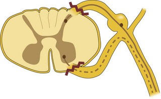
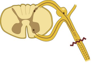
Identification of an avulsed nerve root is important for several reason. Nerve root avulsions are preganglionic lesions. These types of lesions and do not lend themselves to surgical repair (Figure 7.1); however, postganglionic lesions are more amenable to surgical repair (Figure 7.2). The prognosis for recovery of neurological function is unfavorable in preganglionic lesions. When primary repair of the nerve root is not feasible, other surgical options may be available. Preoperative determination that an is present allows the surgeon to determine if surgery is beneficial and to develop a surgical strategy if indicated.


EDX studies can identify neurapraxic lesions. Neurapraxia is presumed when one observes preserved normal motor and sensory nerve conduction distal to the site of injury without denervation on needle electromyography (EMG) of the relevant innervated muscles. However, reduced or absent voluntary motor units may be seen in the muscles. If a child has normal EDX findings but presents with motor and sensory deficits, the electromyographer must be certain to study the clinically involved nerves and muscles to avoid overlooking a more serious injury. Neurapraxic lesions should resolve spontaneously within a period of days to a few weeks. If neurological recovery does not progress as expected, further electrodiagnostic evaluation is warranted.


EDX studies can identify but cannot distinguish axonotmesis from neurotmesis. In neurotmesis in which complete disruption of the neural structure has occurred, surgery leads to the best outcome. However, if axonotmesis is the predominant lesion, axonal re-growth is presumed and surgical nerve repair may or may not result in the best outcome.


In short, EDX studies can help to identify the type, extent, and location of the lesion(s) contributing to neurological deficits in NBPP patients. However, the diagnostic dilemma revolves around what specific EDX tests should one use, the clinical and the specificity and sensitivity of the data obtained.


Nerve conduction studies


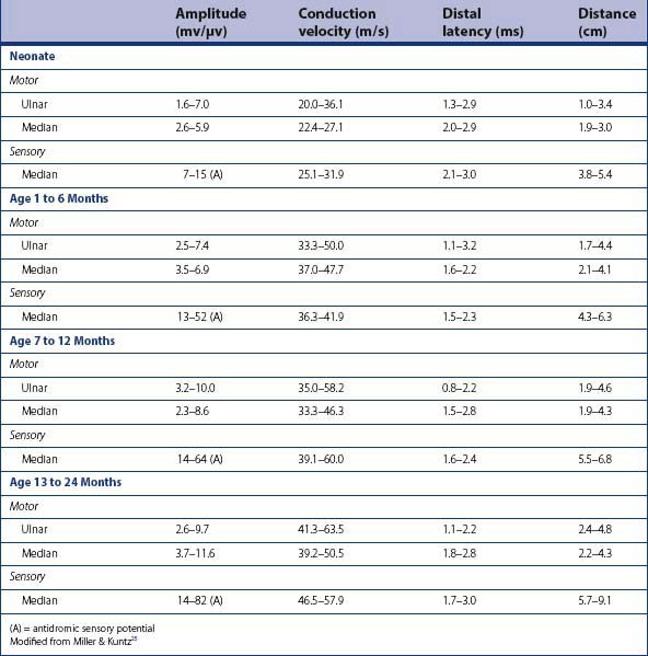
The principles of performing nerve conduction studies (NCS) in the infant and young child are similar to that for the adult patient (refer to Chapter 17) (Figure 7. 3). Due to the infant’s age and size, appropriate modifications must be made (Chart 7.1). It is commonly accepted that the normal values in NCS vary with age (Table 7.1). Motor conduction velocities in newborns are approximately half that of adults (Chart 7.2). Some challenges of NCS in infants exist, such as the difficulty in quantifying axonal loss, and correlating the extent of axonal loss with future recovery.


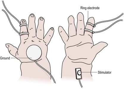
Chart 7.1 Performing nerve conduction studies in infants


< div class='tao-gold-member'>

Stay updated, free articles. Join our Telegram channel

Jul 11, 2016 | Posted by in PEDIATRICS | Comments Off on The role of electrodiagnosis in infants with brachial plexus palsies

Full access? Get Clinical Tree

Get Clinical Tree app for offline access