Chapter 88 The Newborn Infant (See Also Chapter 7)
88.1 History in Neonatal Pediatrics
The perinatal history should include the following information:
88.2 Physical Examination of the Newborn Infant
Many physical and behavioral characteristics of a normal newborn infant are described in Chapters 7 and 584.
General Appearance
Physical activity may be absent during normal sleep, or it may be decreased by the effects of illness or drugs; an infant may be either lying with the extremities motionless, to conserve energy for the effort of difficult breathing, or vigorously crying, with accompanying activity of the arms and legs. Both active and passive muscle tone and any unusual posture should be noted. Coarse, tremulous movements with ankle or jaw myoclonus are more common and less significant in newborn infants than at any other age. Such movements tend to occur when an infant is active, whereas convulsive twitching usually occurs in a quiet state. Edema may produce a superficial appearance of good nutrition. Pitting after applied pressure may or may not be noted, but the skin of the fingers and toes lacks the normal fine wrinkles when filled with fluid. Edema of the eyelids commonly results from irritation caused by the administration of silver nitrate. Generalized edema may occur with prematurity, hypoproteinemia secondary to severe erythroblastosis fetalis, nonimmune hydrops, congenital nephrosis, Hurler syndrome, and from unknown causes. Localized edema suggests a congenital malformation of the lymphatic system; when confined to one or more extremities of a female infant, it may be the initial sign of Turner syndrome (Chapters 76 and 580).
Skin
The vernix and common transitory macular capillary hemangiomas of the eyelids and neck are described in Chapter 639. Cavernous hemangiomas are deeper, blue masses that, if large, may trap platelets and produce disseminated intravascular coagulation or interfere with local organ function. Scattered petechiae may be seen on the presenting part (usually the scalp or face) after a difficult delivery. Slate-blue, well-demarcated areas of pigmentation are seen over the buttocks, back, and sometimes other parts of the body in more than 50% of black, Native American, and Asian infants and occasionally in white ones. These benign patches have no known anthropologic significance despite their name, Mongolian spots; they tend to disappear within the 1st year. The vernix, skin, and especially the cord may be stained brownish yellow if the amniotic fluid has been colored by the passage of meconium during or before birth.
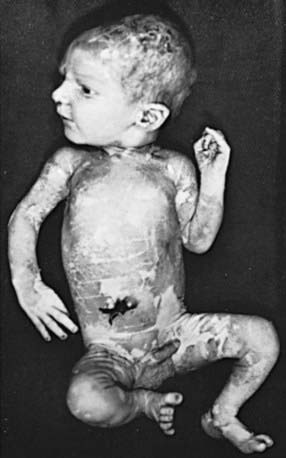
The skin of premature infants is thin and delicate and tends to be deep red; in extremely premature infants, the skin appears almost gelatinous and translucent. Fine, soft, immature hair, lanugo, frequently covers the scalp and brow and may also cover the face of premature infants. Lanugo has usually been lost or replaced by vellus hair in term infants. Tufts of hair over the lumbosacral spine suggest an underlying abnormality such as occult spina bifida, a sinus tract, or a tumor. The nails are rudimentary in very premature infants, but they may protrude beyond the fingertips in infants born past term. Post-term infants may have a peeling, parchment-like skin (Fig. 88-1), a severe degree of which suggests ichthyosis congenita (Chapter 650).
In many neonates, small, white papules on an erythematous base develop 1-3 days after birth. This benign rash, erythema toxicum, persists for as long as 1 wk, contains eosinophils, and is usually distributed on the face, trunk, and extremities (Chapter 639). Pustular melanosis, a benign lesion seen predominantly in black neonates, contains neutrophils and is present at birth as a vesiculopustular eruption around the chin, neck, back, extremities, and palms or soles; it lasts 2-3 days. Both lesions need to be distinguished from more dangerous vesicular eruptions such as herpes simplex (Chapter 244) and staphylococcal disease of the skin (Chapter 174.1).
Skull
The head circumference of all infants should be charted. The skull may be molded, particularly if the infant is the first-born and if the head has been engaged in the pelvic canal for a considerable time. The parietal bones tend to override the occipital and frontal bones. The head of an infant born by cesarean section or from a breech presentation is characterized by its roundness. The suture lines and the size and fullness of the anterior and posterior fontanels should be determined digitally by palpation. Premature fusion of sutures (cranial synostosis) is identified as a hard nonmovable ridge over the suture and an abnormally shaped skull. Great variation in the size of the fontanels exists at birth; if small, the anterior fontanel usually tends to enlarge during the 1st few months of life. The persistence of excessively large anterior (normal, 20 ± 10 mm) and posterior fontanels has been associated with several disorders (Table 88-1). Persistently small fontanels suggest microcephaly, craniosynostosis, congenital hyperthyroidism, or wormian bones; presence of a 3rd fontanel suggests trisomy 21 but is seen in preterm infants. Soft areas (craniotabes) are occasionally found in the parietal bones at the vertex near the sagittal suture; they are more common in premature infants and in infants who have been exposed to uterine compression. Though such soft areas are usually insignificant, their possible pathologic cause should be investigated if they persist. Soft areas in the occipital region suggest the irregular calcification and wormian bone formation associated with osteogenesis imperfecta, cleidocranial dysostosis, lacunar skull, cretinism, and, occasionally, Down syndrome. Transillumination of an abnormal skull in a dark room followed by ultrasound or computed tomography will rule out hydranencephaly and hydrocephaly (Chapter 585). An excessively large head (megalencephaly) suggests hydrocephaly, storage disease, achondroplasia, cerebral gigantism, neurocutaneous syndromes, or inborn errors of metabolism, or may be familial. The skull of a premature infant may suggest hydrocephaly because of the relatively larger brain growth in comparison with growth of other organs. Depression of the skull (indentation, fracture, pingpong ball deformity) is usually of prenatal onset and due to prolonged focal pressure by the bony pelvis. Atrophic or alopecic scalp areas may represent aplasia cutis congenita, which may be sporadic or autosomal dominant or associated with trisomy 13, chromosome 4 deletion, or Johanson-Blizzard syndrome. Deformational plagiocephaly may be due to in utero positioning forces on the skull and manifests as an asymmetric skull and face with ear malalignment. It is associated with torticollis and vertex positioning. Any significant and persistent abnormality in shape or size of the skull should be evaluated by cranial CT.
Face
The general appearance of the face should be noted with regard to dysmorphic features, such as epicanthal folds, widely or narrowly spaced eyes, microphthalmos, asymmetry, long philtrum, and low-set ears, which are often associated with congenital syndromes. The face may be asymmetric as a result of a 7th nerve palsy, hypoplasia of the depressor muscle at the angle of the mouth, or an abnormal fetal posture (Chapter 102); when the jaw has been held against a shoulder or an extremity during the intrauterine period, the mandible may deviate strikingly from the midline. Symmetric facial palsy suggests absence or hypoplasia of the 7th nerve nucleus (Möbius syndrome).
Eyes
The eyes often open spontaneously if the infant is held up and tipped gently forward and backward. This maneuver, a result of labyrinthine and neck reflexes, is more successful for inspecting the eyes than is forcing the lids apart. Conjunctival and retinal hemorrhages are usually benign. Retinal hemorrhages are more common with vacuum-assisted deliveries (75%) than after cesarean section (7%). They resolve in most infants by 2 wk of age (85%) and in all infants by 4 wk. Pupillary reflexes are present after 28-30 wk of gestation. The iris should be inspected for colobomas and heterochromia. A cornea >1cm in diameter in a term infant (with photophobia and tearing) suggests congenital glaucoma and requires prompt ophthalmologic consultation. The presence of bilateral red reflexes suggests the absence of cataracts and intraocular pathology (Chapters 611, 619–625). Leukokoria (white pupillary reflex) suggests cataracts, tumor, chorioretinitis, retinopathy of prematurity, or a persistent hyperplastic primary vitreous and warrants an immediate ophthalmologic consultation.
Mouth
A normal mouth may rarely have precocious dentition, with natal (present at birth) or neonatal (eruption after birth) teeth in the lower incisor position or aberrantly placed; these teeth are shed before the deciduous ones erupt (Chapter 299). Alternatively, such teeth occur in Ellis-van Creveld, Hallermann-Streiff, and other syndromes. Extraction is not usually indicated. Premature eruption of deciduous teeth is even more unusual. The soft and hard palate should be inspected and palpated for a complete or submucosal cleft, and the contour noted if the arch is excessively high or the uvula is bifid. On the hard palate on either side of the raphe, there may be temporary accumulations of epithelial cells called Epstein pearls. Retention cysts of similar appearance may also be seen on the gums. Both disappear spontaneously, usually within a few weeks of birth. Clusters of small white or yellow follicles or ulcers on erythematous bases may be found on the anterior tonsillar pillars, most frequently on the 2nd or 3rd day of life. Of unknown cause, they clear without treatment in 2-4 days.
Neck
The neck appears relatively short. Abnormalities are not common but include goiter, cystic hygroma, branchial cleft rests, teratoma, hemangioma, and lesions of the sternocleidomastoid muscle that are presumably traumatic or due to a fixed positioning in utero that produces either a hematoma or fibrosis, respectively. Congenital torticollis causes the head to turn toward and the face to turn away from the affected side. Plagiocephaly, facial asymmetry, and hemihypoplasia may develop if it is untreated (Chapter 672.1). Redundant skin or webbing in a female infant suggests intrauterine lymphedema and Turner syndrome (Chapters 76 and 580). Both clavicles should be palpated for fractures.


