The Neck

Chapter 672 The Neck


672.1 Torticollis


Torticollis is a symptom rather than a diagnosis, and describes the clinical findings of tilting of the head to the right or left side in combination with rotation of the head to the opposite side. Congenital muscular torticollis (CMT) is the most common etiology, but a variety of other conditions result in torticollis, and a detailed workup is often required to rule out other diagnoses in patients who lack the characteristic features of CMT (approximately 20%) (Fig. 672-1). The differential diagnosis includes trauma (clavicle fracture or brachial plexopathy), tumors or malformations of the central nervous system, ocular disorders, congenital bony abnormalities (Klippel-Feil syndrome), inflammatory conditions, and other diagnoses such as atlantoaxial rotatory displacement or Sandifer syndrome (Table 672-1).
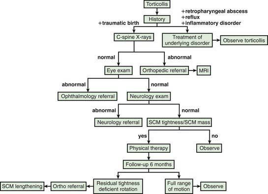


The majority of cases discovered within the first few months of life represent congenital muscular torticollis (CMT), which occurs in up to 1/250 live births. In newborns, other diagnoses include clavicle fracture and/or brachial plexopathy. Although the etiology of CMT remains unknown, current evidence (muscle biopsies and MRI studies showing fibrosis) supports in utero muscular compression and/or stretch, possibly resulting in localized ischemia and an intramuscular compartment syndrome. CMT is more common in firstborn children and following difficult births.


A contracture of the left sternocleidomastoid muscle results in tilt of the head to the left and rotation to the right, and vice versa. A fibrotic mass is palpable within the substance of the sternocleidomastoid muscle in approximately 50% of patients, and it usually disappears within the first months of life, often being replaced by a fibrous band. CMT may be seen as part of a molded baby syndrome, occurring in combination with other findings thought to relate to intrauterine mechanical deformation such as developmental hip dysplasia, plagiocephaly, facial asymmetry, and foot deformities such as metatarsus adductus. A prospective study of 102 consecutive newborns demonstrated morphologic “asymmetries” in 73%, including torticollis (16%), mandibular asymmetry (13%), facial asymmetry (42%), and skull asymmetry (61%). Facial findings associated with CMT include flattening on the affected side, recessed eyebrow and zygoma, and inferior orbital displacement. There is also evidence to suggest that persistent sternocleidomastoid contracture can result in progressive deformation; although morphologic abnormalities of the cranium and cranial base may be observed in infancy, facial bone asymmetry develops at or later than 5 yr of age. Hip dysplasia occurs in ∼3-9% of patients with CMT, and although guidelines for screening in patients with a normal hip examination have not been established, consideration should be given to obtaining either an ultrasound scan (1 mo of age) or a plain radiograph of the hip (4-5 mo of age). A delay in achieving early developmental milestones has been reported in babies with CMT, but this is most likely explained by a decrease in prone positioning while awake.


Torticollis can also result from congenital vertebral anomalies (including Klippel-Feil syndrome). Although there are no formal guidelines for when to order plain radiographs to rule out an underlying congenital osseous abnormality, radiographs of the cervical spine are suggested when the typical clinical features associated with congenital muscular torticollis are absent or if the deformity does not respond to treatment. This recommendation is supported by a study in which congenital vertebral anomalies were identified on screening radiographs in only 4 of 502 infants with torticollis in the absence of birth trauma; radiographic findings that would serve as a contraindication to a stretching program were only identified in a single patient.


The treatment of congenital muscular torticollis involves stretching, stimulation, and positioning measures, often supervised by a physical therapist. Resolution should be achieved in approximately 95% of cases, especially when the treatment is started during the first 4 mo of life. Intramuscular injection of botulinum toxin A (Botox) into the sternocleidomastoid may be considered in resistant cases of CMT, although further study is required to determine the effectiveness and indications for this modality. Complications include transient dysphagia and neck weakness.


For patients with a late diagnosis or those in whom the stretching program has failed to correct the deformity, surgical release of the sternocleidomastoid is considered. The optimal timing for surgery continues to be debated. Some authors have suggested that the procedure be performed at 12-18 mo of age to facilitate remodeling of craniofacial molding abnormalities, due to the greater growth potential in the younger patients. Others have suggested that outcomes are improved when the surgery is delayed until later, even to school age. Motion can be improved following surgical release even in teenagers. One study compared a cohort from 1-4 yr of age with another from 5-16 yr of age and concluded that although there were no significant differences in craniofacial asymmetry, residual contracture, and subjective measures, the older age group had less surgical scarring and head tilt. Surgical management results in adequate function and acceptable cosmesis in >90% of patients. With early diagnosis and treatment, surgery should be required in only a minority of cases.


The evaluation of torticollis becomes more complex when the typical findings associated with CMT are absent, the usual clinical response is not observed, or the deformity occurs at a later age. A careful history and physical examination is essential, supplemented by additional imaging studies and often consultation with an ophthalmologist, neurologist, or other specialists. Plain radiographs should be obtained to rule out an underlying congenital osseous abnormality, and an MRI of the brain and cervical spine are required in many cases to rule out a tumor (posterior fossa or brainstem) or a developmental condition such as a Chiari I malformation and/or syringomyelia.


Torticollis can result from congenital vertebral anomalies or congenital scoliosis, and progressive deformities can require surgical stabilization. Ocular torticollis can result from strabismus (weakness of the 4th cranial nerve) or a superior oblique muscle palsy. Sandifer’s syndrome describes torticollis in association with gastroesophageal reflux. Atlantoaxial rotatory displacement represents a spectrum of rotational malalignment (subluxation to dislocation) between the atlas (C1) and the axis (C2), and may best be described as pathologic stickiness in the arc of joint motion. The malalignment may initially be reducible, but after weeks to months the deformity becomes fixed and irreducible. Thus, prompt diagnosis and treatment are essential.


A variety of conditions can lead to rotatory displacement, including infection or inflammation of the tissues of the upper airway, neck, and/or pharynx (Grisel syndrome). Traumatic injuries (usually minor) have been associated, and rotatory displacement occasionally complicates surgical procedures in the oropharynx, ear, or nose. The diagnosis is confirmed with a CT scan in which axial images are obtained from the occiput through C2 in neutral alignment and with maximal rotation to the right and to the left. The images essentially define the motion curve between the 2 vertebrae and determine whether any rotational malalignment is reducible, partially reducible, or fixed. Rotatory fixation exists when the relationship between C1 and C2 remains constant through the arc of motion.


The treatment varies based on the underlying pathology and the chronicity of symptoms. If the patient is seen within a few days of the onset of symptoms, then a trial of analgesics and a soft collar may be attempted. Patients with symptoms for >1 wk are often admitted to the hospital for analgesia, muscle relaxants, and a period of cervical traction. If this fails to restore normal anatomy and motion, then halo traction may be attempted. The amount of force transmitted is limited to 5-8 lb with cervical traction, due to pressure on the mandible. Much greater traction weights may be applied when a halo or Gardner Wells tongs are applied. If the malalignment is corrected and full cervical motion is restored, patients are typically immobilized for at least 6 wk in a halo vest. A pinless halo has been employed in some centers to immobilize these patients, because the device does not require pins to be placed into the skull and is better tolerated. Patients who fail to respond to traction, typically those with a fixed deformity, and those in whom the malalignment has recurred, may require a posterior atlantoaxial fusion to stabilize the articulation.


Paroxysmal torticollis of infancy

Stay updated, free articles. Join our Telegram channel

Jun 18, 2016 | Posted by in PEDIATRICS | Comments Off on The Neck

Full access? Get Clinical Tree

Get Clinical Tree app for offline access