Chapter 462 The Inherited Pancytopenias
Pancytopenia refers to a reduction below normal values of all 3 peripheral blood lineages: leukocytes, platelets, and erythrocytes. Pancytopenia requires microscopic examination of a bone marrow biopsy specimen and a marrow aspirate to assess overall cellularity and morphology. There are 3 general categories of pancytopenia depending on the marrow findings.
Inherited (“constitutional”) pancytopenia is defined as a decrease in marrow production of the 3 major hematopoietic lineages that occurs on an inherited basis, resulting in anemia, neutropenia, and thrombocytopenia. Any of these conditions (Table 462-1) can be transmitted as a simple mendelian disorder by mutant genes with inherited patterns of autosomal dominant, autosomal recessive, or X-linked types. Modifying genes and acquired factors may also be operative. Inherited pancytopenias account for approximately 30% of cases of pediatric marrow failure. Fanconi anemia is the most common of these disorders.
Fanconi Anemia
Etiology and Epidemiology
Fanconi anemia (FA) is primarily inherited in an autosomal recessive manner (one uncommon form is X-linked recessive). It occurs in all racial and ethnic groups. At presentation, patients with FA may have: (1) typical physical anomalies but normal hematologic findings; (2) normal physical features but abnormal hematologic findings; or (3) physical anomalies and abnormal hematologic findings, which constitute the classic phenotype (39% of cases). There can be sibling discordance in clinical and hematologic findings, even in affected monozygotic twins. Approximately 75% of patients are 3-14 yr of age at the time of diagnosis.
Pathology
Patients have abnormal chromosome fragility, which is seen in metaphase preparations of peripheral blood lymphocytes cultured with phytohemagglutinin and enhanced by adding clastogenic agents such as diepoxybutane (DEB) and mitomycin C. Cell fusion of FA cells with normal cells or with cells from some unrelated patients with FA produces a corrective effect on chromosomal fragility, a process called complementation. This phenomenon allows subtyping of cases of FA into discrete complementation groups. Fourteen separate complementation groups have been identified, and 14 mutant FA (FANC) genes have been cloned so far (A, B, C, D1/BRCA2, D2, E, F, G, I, J, L, M, N, and O) all prefixed with FANC, e.g. FANCA, FANCB and so on); FANCD1 is identical to the breast cancer susceptibility protein BRCA2. The protein products of wild-type FANC genes are involved in the DNA damage recognition and repair biochemical pathways. Therefore, mutant gene proteins lead to genomic instability, chromosome fragility, and FA. An inability of FA cells to remove oxygen-free radicals, resulting in oxidative damage, is a contributing factor in the pathogenesis. Additional factors are also operative. Leukocyte telomere length is significantly shortened but telomerase activity is increased, suggesting a high proliferative rate of marrow progenitors that ultimately leads to their premature senescence. Increased marrow cell apoptosis occurs and is mediated by Fas, a membrane glycoprotein receptor containing an integral death domain. A consistent finding is diminished cellular interleukin-6 production along with markedly heightened tumor necrosis factor-α generation.
Clinical Manifestations
The most common anomaly in FA is hyperpigmentation of the trunk, neck, and intertriginous areas, as well as café-au-lait spots and vitiligo, alone or in combination (Fig. 462-1 and Table 462-2). Half the patients have short stature. Growth failure may be associated with abnormal growth hormone secretion or with hypothyroidism. Absence of radii and thumbs that are hypoplastic, supernumerary, bifid, or absent are common. Anomalies of the feet, congenital hip dislocation, and leg abnormalities are seen. A male patient with FA may have an underdeveloped penis; undescended, atrophic, or absence of the testes; and hypospadias or phimosis. Females can have malformations of the vagina, uterus, and ovary. Many patients have a FA “facies,” including microcephaly, small eyes, epicanthal folds, and abnormal shape, size, or positioning of the ears (see Fig. 462-1). Ectopic, pelvic, or horseshoe kidneys are detected by imaging and may show other organs as duplicated, hypoplastic, dysplastic, or absent kidneys. Cardiovascular and gastrointestinal malformations also occur. Approximately 10% of patients with FA are cognitively delayed.

Figure 462-1 A 3 yr old boy with Fanconi anemia who exhibits several classic phenotype features. A, Front view. B, Face. C, Hands. D, Back right shoulder. The features to be noted include short stature, dislocated hips, microcephaly, a broad nasal base, epicanthal folds, micrognathia, thumbs attached by a thread, and café-au-lait spots with hypopigmented areas beneath.
(From Nathan DC, Orkin SH, Ginsburg D, et al, editors: Nathan and Oski’s hematology of infancy and childhood, ed 6, vol I, Philadelphia, 2003, WB Saunders, p 285.)
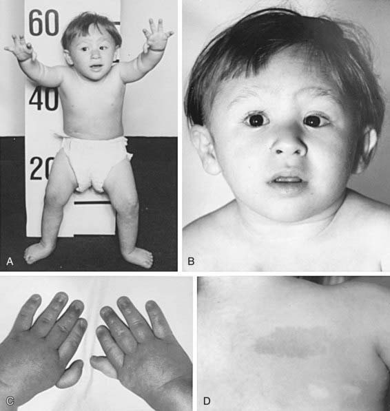
Table 462-2 CHARACTERISTIC PHYSICAL ANOMALIES IN FANCONI ANEMIA
ANOMALY | APPROXIMATE FREQUENCY (% OF PATIENTS) |
---|---|
Skin pigment changes ± café-au-lait spots | 55 |
Short stature | 51 |
Upper limb abnormalities (thumbs, hands, radii, ulnas) | 43 |
Hypogonadal and genital changes (mostly male) | 35 |
Other skeletal findings (head/face, neck, spine) | 30 |
Eye/lid/epicanthal fold anomalies | 23 |
Renal malformations | 21 |
Gastrointestinal/cardiopulmonary malformations | 11 |
Hip, leg, foot, toe abnormalities | 10 |
Ear anomalies (external and internal), deafness | 9 |
Laboratory Findings
Marrow failure usually ensues in the 1st decade of life. Thrombocytopenia often appears initially, with subsequent onset of granulocytopenia and then macrocytic anemia. Severe aplasia develops in most cases, but its full expression is variable and evolves over a period of months to years. The marrow becomes progressively hypocellular and fatty, like that in severe acquired aplastic anemia. Chromosome fragility is indicated by spontaneously occurring chromatid breaks, rearrangements, gaps, endoreduplications, and chromatid exchanges in blood lymphocytes cultured with phytohemagglutinin as well as in cultured skin fibroblasts, underscoring the constitutional nature of the disorder. With addition of DEB, fragility is strikingly enhanced in lymphocyte cultures of patients with FA in comparison with those of controls. For prenatal diagnosis, abnormal chromosome breakage can be tested for in amniotic fluid cells or in tissue from a chorionic villus biopsy.
Complications
A major feature of the phenotype of FA is the propensity for cancer. The most frequent solid tumors are squamous cell carcinomas of the head, neck, and upper esophagus, followed by carcinomas of the vulva and/or anus, cervix, and lower esophagus. Human papilloma virus is suspected in the pathogenesis. Some patients experience oral cancer after bone marrow transplantation. Benign and malignant liver tumors occur (adenomas, hepatomas) and are usually associated with androgen therapy for aplastic anemia. Androgens are also implicated in the etiology of peliosis hepatis (blood-filled hepatic sinusoids). Peliosis hepatis is reversible when androgen therapy is discontinued, and tumors may regress. Approximately 15% of patients with FA are at risk for acute leukemia or MDS.
Diagnosis
FA should be considered in all children and young adults with unexplained cytopenias. Abnormal hematologic findings and characteristic physical anomalies suggest the diagnosis, which is confirmed with a lymphocyte chromosomal breakage study using DEB. No other inherited pancytopenia is associated with an abnormal DEB chromosomal breakage study result. Ten percent to 15% of patients with suspected FA have “somatic mosaicism” and do not show abnormal lymphocyte chromosomal fragility because of mixed populations of somatic cells, some with 2 abnormal alleles and some with 1. Testing on skin fibroblast cultures instead of lymphocytes confirms the diagnosis.
Most patients have stable elevations of serum α-fetoprotein expressed constitutively, independent of liver complications or androgen therapy. The laboratory measurement of serum α-fetoprotein can be used as a rapid screening diagnostic test.
Specialized laboratories can perform an accurate diagnostic and mutant gene subtyping assay, whereby patient lymphocytes or fibroblasts are studied after exposure to mitomycin C or radiation by immunoblotting for FANCD2. Alternatively, wild-type FANC genes can be transvected into patient T cells by means of retroviral vectors, and if a specific wild-type FANC gene corrects the abnormal T-cell chromosome fragility, the specific mutant gene is deduced.
Treatment
A hematologist and a multidisciplinary team should supervise patients with FA. If the hematologic findings are stable and there are no transfusion requirements, observation is indicated. Subspecialty consultations for anomalies and disabilities can be arranged during this interval. If growth velocity is below expectations, endocrine evaluation is needed to identify growth hormone deficiency or hypothyroidism. Screening for glucose intolerance and hyperinsulinemia should be performed annually or biannually, depending on the degree of hyperglycemia found on initial testing. Blood counts should be performed every 1-3 mo; bone marrow aspiration and biopsy are indicated annually for leukemia and MDS surveillance by means of morphology and cytogenetics. Patients should be assessed for solid tumors at least annually. Beginning at menarche, female patients should be screened annually for gynecologic cancer. Administration of human papilloma virus quadrivalent vaccine to prevent squamous cell carcinoma will likely become a standard intervention.
Hematopoietic stem cell transplantation (HSCT; Chapter 129) is the only curative therapy for the hematologic abnormalities. Patients with FA <10 yr old who undergo transplantation using an HLA–identical sibling donor have a survival rate >80%. Survival rates are lower for patients undergoing the procedure when >10 yr. Preparative regimens are continuously evaluated, refined, and improved worldwide. For patients who do not have a matched sibling donor, a search for a matched unrelated donor (including a search of umbilical cord blood banks) might be initiated. Because of the heightened graft vs host response in patients with FA, the survival and cure rates have not been as good as those for matched sibling donor HSCT (≈ 30% survival). Molecular technology has led to preimplantation genetic diagnosis on parent-derived blastomeres to find an HLA-matched sibling donor without FA.
Androgens produce a response in 50% of patients, heralded by reticulocytosis and a rise in hemoglobin within 1-2 mo. White blood cell counts may increase next, followed by platelet counts, but it may take many months to achieve the maximum response. When the response plateaus, androgen dosage can be slowly tapered but not stopped entirely. Oral oxymetholone is used most frequently once a day. Low-dose prednisone orally every 2nd day may be added to counter androgen-induced growth acceleration and prevent thrombocytopenic bleeding by promoting vascular stability. In many patients who are taking androgens, the disease becomes refractory as marrow failure progresses. Potential side effects include masculinization, elevated hepatic enzymes, cholestasis, peliosis hepatis, and liver tumors. Screening for these changes should be performed serially.
The premise for gene therapy in FA is based on the assumption that corrected hematopoietic cells offer a growth advantage. Attempts at gene therapy have been disappointing, possibly because of the type of vector but also because of the chromosomal fragility and impaired proliferative function of the hematopoietic progenitors. Encouraging preclinical data from studies using lentiviral vectors offer hope that gene therapy will be a safe and effective treatment for FA. Transposons are nonviral vectors that have been used successfully for gene delivery in murine models and may hold promise for use in humans.

Full access? Get Clinical Tree

