The Hip

Chapter 670 The Hip


The hip joint is a pivotal joint of the lower extremity, and its functional demands require both stability and flexibility. Anatomically, the hip joint is a ball-and-socket articulation between the femoral head and acetabulum.


Growth and Development


The hip joint begins to develop at about the 7th week of gestation, when a cleft appears in the mesenchyme of the primitive limb bud. These precartilaginous cells differentiate into a fully formed cartilaginous femoral head and acetabulum by the 11th week of gestation (Chapter 6.1). At birth, the neonatal acetabulum is completely composed of cartilage, with a thin rim of fibrocartilage called the labrum.


The very cellular hyaline cartilage of the acetabulum is continuous with the triradiate cartilages, which divide and interconnect the three osseous components of the pelvis (the ilium, ischium, and pubis). The concave shape of the hip joint is determined by the presence of a spherical femoral head.


Several factors determine acetabular depth, including interstitial growth within the acetabular cartilage, appositional growth under the perichondrium, and growth of adjacent bones (the ilium, ischium, and pubis). In the neonate, the entire proximal femur is a cartilaginous structure, which includes the femoral head and the greater and lesser trochanters. The three main growth areas are the physeal plate, the growth plate of the greater trochanter, and the femoral neck isthmus. Between the 4th and 7th mo of life, the proximal femoral ossification center (in the center of the femoral head) appears. This ossification center continues to enlarge, along with its cartilaginous anlage, until adult life, when only a thin layer of articular cartilage remains. During this period of growth, the thickness of the cartilage surrounding this bony nucleus gradually decreases, as does the thickness of the acetabular cartilage. The growth of the proximal femur is affected by muscle pull, the forces transmitted across the hip joint with weight bearing, normal joint nutrition, circulation, and muscle tone. Alterations in these factors can cause profound changes in development of the proximal femur.


Vascular Supply


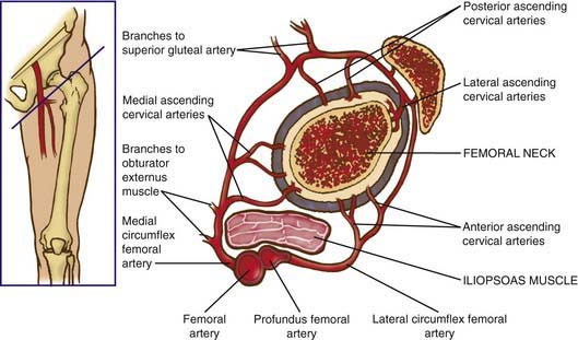
The blood supply to the capital femoral epiphysis is complex and changes with growth of the proximal femur. The proximal femur receives its arterial supply from intraosseous (primarily the medial femoral circumflex artery) and extraosseous vessels (Fig. 670-1). The retinacular vessels (extraosseous) lie on the surface of the femoral neck but are intracapsular because they enter the epiphysis from the periphery. This makes the blood supply vulnerable to damage from septic arthritis, trauma, thrombosis, and other vascular insults. Interruption of this tenuous blood supply can lead to avascular necrosis of the femoral head and permanent deformity of the hip.


670.1 Developmental Dysplasia of the Hip


Developmental dysplasia of the hip (DDH) refers to a spectrum of pathology in the development of the immature hip joint. The original term for the condition, congenital dislocation of the hip, was replaced by the current name to more accurately reflect the variable presentation of the disorder and to encompass mild dysplasias as well as frank dislocations.


Clinical Findings


The Neonate


DDH in the neonate is asymptomatic and must be screened for by specific maneuvers. Physical examination must be carried out with the infant unclothed and placed supine in a warm, comfortable setting on a flat examination table.


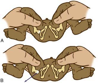
The Barlow provocative maneuver assesses the potential for dislocation of a nondisplaced hip. The examiner adducts the flexed hip and gently pushes the thigh posteriorly in an effort to dislocate the femoral head (Fig. 670-2). In a positive test, the hip is felt to slide out of the acetabulum. As the examiner relaxes the proximal push, the hip can be felt to slip back into the acetabulum.


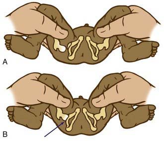
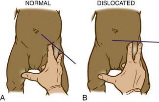
The Ortolani test is the reverse of Barlow test: The examiner attempts to reduce a dislocated hip (Fig. 670-3). The examiner grasps the child’s thigh between the thumb and index finger and, with the 4th and 5th fingers, lifts the greater trochanter while simultaneously abducting the hip. When the test is positive, the femoral head will slip into the socket with a delicate clunk that is palpable but usually not audible. It should be a gentle, nonforced maneuver.


A hip click is the high-pitched sensation (or sound) felt at the very end of abduction during testing for DDH with Barlow and Ortolani maneuvers. Classically, a hip click is differentiated from a hip clunk, which is felt as the hip goes in and out of joint. Hip clicks usually originate in the ligamentum teres or occasionally in the fascia lata or psoas tendon and do not indicate a significant hip abnormality.


The Infant


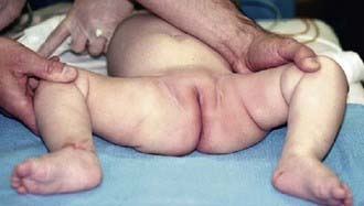
As the baby enters the 2nd and 3rd months of life, the soft tissues begin to tighten and the Ortolani and Barlow tests are no longer reliable. In this age group, the examiner must look for other specific physical findings including limited hip abduction, apparent shortening of the thigh, proximal location of the greater trochanter, asymmetry of the gluteal or thigh folds (Fig. 670-4), and pistoning of the hip. Limitation of abduction is the most reliable sign of a dislocated hip in this age group.


Shortening of the thigh, the Galeazzi sign, is best appreciated by placing both hips in 90 degrees of flexion and comparing the height of the knees, looking for asymmetry (Fig. 670-5). Asymmetry of thigh and gluteal skin folds may be present in 10% of normal infants but suggests DDH. Another helpful test is the Klisic test, in which the examiner places the 3rd finger over the greater trochanter and the index finger of the same hand on the anterior superior iliac spine. In a normal hip, an imaginary line drawn between the two fingers points to the umbilicus. In the dislocated hip, the trochanter is elevated, and the line projects halfway between the umbilicus and the pubis (Fig. 670-6).
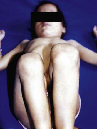


Diagnostic Testing


Ultrasonography


Because it is superior to radiographs for evaluating cartilaginous structures, ultrasonography is the diagnostic modality of choice for DDH before the appearance of the femoral head ossific nucleus (4-6 mo). During the early newborn period (0-4 wk), however, physical examination is preferred over ultrasonography because there is a high incidence of false-positive sonograms in this age group. In addition to elucidating the static relationship of the femur to the acetabulum, ultrasonography provides dynamic information about the stability of the hip joint. The ultrasound examination can be used to monitor acetabular development, particularly of infants in Pavlik harness treatment; this method can minimize the number of radiographs taken and might allow the clinician to detect failure of treatment earlier.


In the Graf technique, the transducer is placed over the greater trochanter, which allows visualization of the ilium, the bony acetabulum, the labrum, and the femoral epiphysis (Fig. 670-7). The angle formed by the line of the ilium and a line tangential to the boney roof of the acetabulum is termed the α angle and represents the depth of the acetabulum. Values >60 degrees are considered normal, and those <60 degrees imply acetabular dysplasia. The β angle is formed by a line drawn tangential to the labrum and the line of the ilium; this represents the cartilaginous roof of the acetabulum. A normal β angle is <55 degrees; as the femoral head subluxates, the β angle increases. Another useful test is to evaluate the position of the center of the head compared to the vertical line of the ilium. If the line of the ilium falls lateral to the center of the head, the epiphysis is considered reduced. If the line falls medial to the center of the head, the epiphysis is undercovered and is either subluxated or dislocated.


Screening for DDH with ultrasound remains controversial. Although routinely performed in Europe, ultrasonographic screening has not been shown to be cost effective in the USA largely because of the cost associated with treating false-positive results. The current recommendations are that every newborn undergo a clinical examination for hip instability. Children who have findings suspicious for DDH should be followed up with ultrasound. Most authors agree that infants with risk factors for DDH (breech position, family history, torticollis) should be screened with ultrasound regardless of the clinical findings.


Radiography


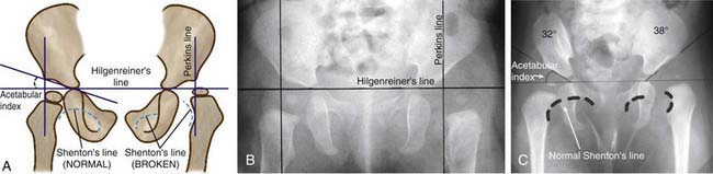
Radiographs are recommended for an infant once the proximal femoral epiphysis ossifies, usually by 4-6 mo. In infants of this age, the radiographs have proved to be more effective, less costly, and less operator dependent than an ultrasound examination. An anteroposterior (AP) view of the pelvis can be interpreted through several classic lines drawn on it (Fig. 670-8).


The Hilgenreiner line is a horizontal line drawn through the top of both triradiate cartilages (the clear area in the depth of the acetabulum). The Perkins line, a vertical line through the most lateral ossified margin of the roof of the acetabulum, is perpendicular to the Hilgenreiner line. The ossific nucleus of the femoral head should be located in the medial lower quadrant of the intersection of these two lines. The Shenton line is a curved line drawn from the medial aspect of the femoral neck to the lower border of the superior pubic ramus. In a child with normal hips, this line is a continuous contour. In a child with hip subluxation or dislocation, this line consists of two separate arcs and therefore is described as “broken.”


The acetabular index is the angle formed between the Hilgenreiner line and a line drawn from the depth of the acetabular socket to the most lateral ossified margin of the roof of the acetabulum. This angle measures the development of the osseous roof of the acetabulum. In the newborn, the acetabular index can be up to 40 degrees; by 4 mo in the normal infant, it should be no more than 30 degrees. In the older child, the center-edge angle

Stay updated, free articles. Join our Telegram channel

Jun 18, 2016 | Posted by in PEDIATRICS | Comments Off on The Hip

Full access? Get Clinical Tree

Get Clinical Tree app for offline access