Fig. 1
Postero-anterior radiograph of a wrist with Grade 3 gymnast wrist . Note the widening of the distal radial physis
The differential diagnosis includes scaphoid impaction syndrome and dorsal impingement syndrome. Scaphoid impaction syndrome is the impaction of the dorsal rim of the scaphoid on the dorsal lip of the radius from forced wrist hyperextension. On physical examination, pain is usually worse dorsally and is accentuated with hyperextension. Radiographs may show an ossicle or a hypertrophic ridge at the dorsal scaphoid rim. In dorsal impingement syndrome , synovitis or capsulitis develops between the dorsal rim of the radius and the carpus. Pain is more localized to the joint rather than to the distal radius. Radiographs may show development of an osteophyte on the dorsal rim of the radius.


Fig. 2
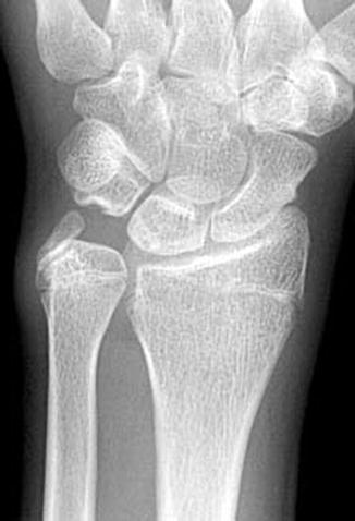
Postero-anterior radiograph of a wrist with stage 3 gymnast wrist . The distal radial physis has closed, yet the ulnar physis is still open, albeit closing. Three millimeter positive ulnar variance is present. Sclerosis is also present in the proximal, ulnar lunate, suggestive of ulnocarpal impaction syndrome
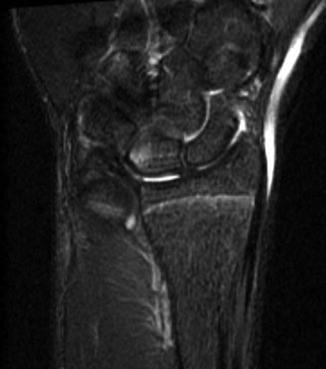
In the older gymnast, the pain is typically worse about the ulnar side of the wrist. If the distal radial physeal stress injury was not recognized or was not treated in a timely fashion, an early physeal closure of the radius occurs, leading to a nonphysiologic ulnar positive wrist (Fig. 2). Patients exhibit signs suggestive of ulnocarpal impaction syndrome : pain over the ulnocarpal joint with ulnar deviation, pain with palpation distal to the ulnar styloid, and pain with stressing the distal radioulnar joint. Plain radiographs typically reveal a positive ulnar variance and cyst formation or sclerosis in the ulnar head or lunotriquetral region (Davis 2010; Gabel 1998). Magnetic resonance imaging (MRI) findings can assist in making the diagnosis. Subchondral bone marrow edema is an indirect sign of chondromalacia and is typically a precursor to radiographic findings. As the syndrome progresses, subchondral sclerosis can appear as areas of low signal intensity on both T1- and T2-weighted images (Fig. 3).


Fig. 3
MRI of the same wrist as seen in Fig. 2. The T2 image shows a hypodense region of the proximal, ulnar lunate, consistent with ulnocarpal impaction syndrome

Fig. 4
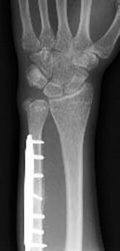
Postoperative postero-anterior radiograph of the same wrist as seen in Fig. 2 following an extra-articular ulnar shortening osteotomy . An oblique osteotomy was performed to permit placement of a lag screw across the osteotomy site. Ulnar variance was restored to neutral
Treatment
The management and prognosis of distal radial stress reaction depend on the stage of the process. Patients are divided into three stages. In stage 1, the diagnosis is made clinically prior to radiographic abnormalities. Management consists of the avoidance of axial compressive loading events. Gradual return to participation can begin once symptoms cease, usually in 2–4 weeks (DiFiori et al. 2006; Rettig 2004). Careful observation during return to gymnastics is crucial because recurrence of pain requires reinstitution of restrictions (DiFiori et al. 2006). Stage 2 demonstrates radiographic distal radial physeal changes without secondary ulnar positive variance. These patients tend to have a poorer prognostic outcome. Return to competition should be delayed for at least 2–4 months (Rettig 2004). DiFiori et al. (2006) recommends that gymnasts in stage 2 refrain from compression loading of the wrist for at least 6 weeks. In individuals with abnormal radiographic findings, repeat radiographs should be performed to determine if the distal radial physeal injury is resolving (DiFiori et al. 2006). Once symptoms have resolved (full, painless wrist range of motion) and radiographs demonstrate resolution of the distal radial physeal injury, then training may resume. Rehabilitation of the extremity should include evaluation of overall limb alignment, laxity, and strengthening and proprioception (DiFiori et al. 2006). If the gymnast is experiencing pain with non-gymnastic activities, then bracing or cast immobilization should be initiated (DiFiori et al. 2006). However, the use of bracing to speed recovery is not well studied.
Stage 3 represents a relatively late presentation, with secondary shortening of the radius and a relative positive ulnar variance. It is this group of gymnasts that are susceptible to ulnar abutment pain (Davis 2010; DiFiori et al. 2006; Rettig 2004). Nonoperative treatment consists of rest, intermittent immobilization, nonsteroidal antiinflammatory drugs, and modification of activities (avoidance of ulnar deviation maneuvers) (Gabel 1998; Sachar 2008). Corticosteroid injection into the ulnocarpal joint can be both therapeutic and diagnostic. If symptoms do not resolve within 6 weeks, then an MRI should be ordered to evaluate the TFCC and intracarpal ligaments. The goal of surgical intervention for ulnar carpal abutment syndrome should be to unload or decompress the ulnar side of the wrist joint (Gabel 1998,; Sachar 2008). Two options are available: ulnar-shortening osteotomy and an open or arthroscopic wafer procedure. The arthroscopic wafer procedure uses a power burr to remove approximately 2 mm of the ulnar head through the TFCC tear (Sachar 2008; Bernstein et al. 2004). Concern for damaging the distal ulnar physis makes this a less desirable procedure in the skeletally immature athlete. The ulnar-shortening osteotomy is performed at the junction of the distal and middle third of the ulna. Typically an oblique osteotomy is created using cutting guides to increase precision (Sachar 2008). The goal of the surgery is to return the ulna to a neutral height compared to the radius (Fig. 4).
Technique: Ulnar Shortening Osteotomy
A non-sterile upper arm tourniquet is placed over cast padding. The arm is exsanguinated and the tourniquet inflated. A 7–10 cm incision is made over the subcutaneous border of the ulnar, beginning 1 cm proximal to the ulnar styloid (in the region of the ulnar neck) and continuing proximally. Care is taken to protect the dorsal sensory branch of the ulnar nerve, which may be visualized in the distal extent of the wound. The plane of dissection is between the flexor carpi ulnaris and the extensor carpi ulnaris muscles and tendons.
The plate can be placed palmarly, dorsally, or ulnarly. The type and size of the cutting jig often determines the best location for the plate. Care should also be taken to place the plate on a flat surface of the bone. The amount of bone to be removed is determined on preoperative X-rays, comparing the involved wrist to the contralateral side. The goal of surgery is to make the involved side at least ulna neutral. Radiographs should be done in neutral forearm rotation to standardize the two sides. Once the osteotomy is performed and the bone disc removed, the osteotomy is compressed. How this occurs is dependent on the system used. A lag screw is placed across the oblique osteotomy.
If the ulnar physis is still open, an epiphysiodesis of the distal ulna is done. The incision is extended distally to the site of the physis. Care is taken to protect the dorsal sensory branch of the ulnar nerve, which may be visualized in the distal extent of the wound. The physis is identified under fluoroscopic guidance. A guide wire for a 2.5 mm cannulated drill is inserted into the physis in an ulnar to radial fashion. The drill bit is then placed over the wire and drilled to but not through the radial cortex. Curved curettes are then used to remove more of the physis from the dorsal and ulnar regions thru this drill hole. The outer regions of the physis are left intact. Bone chips from the ulnar osteotomy may then be placed into the defect in the epiphysiodesis site.
The tourniquet is deflated and hemostasis is obtained. Subsequently, the fascia is repaired above the plate and the skin is closed. The patient is placed in a well padded, long arm splint. On the first postoperative visit, the splint is removed and the incision is assessed for healing. Whether the arm is placed into a long arm or short arm cast is based on the attending surgeon’s preference and the ability of the child to curtail his/her activities. The arm should be protected until radiographic healing is obtained, usually in 6–8 weeks. Rehabilitation begins at this time. Return to sports is usually at 3 months from surgery.
Stay updated, free articles. Join our Telegram channel

Full access? Get Clinical Tree


