The child of uncertain sex

19.3 The child of uncertain sex


Disorders of sexual development are congenital conditions in which the development of chromosomal, gonadal or phenotypic sex is atypical. This includes infants with a genital appearance that does not permit gender assignment:


The birth of a child with a disorder of sexual development presents a psychosocial crisis for the family and may indicate an underlying medical condition such as congenital adrenal hyperplasia, which could be life-threatening if undiagnosed and untreated.


Disorders of sexual development (DSDs) are rare (1 in 4500), often complex, and always require urgent expert consultation. Optimal care requires an experienced multidisciplinary team.


Normal sexual differentiation


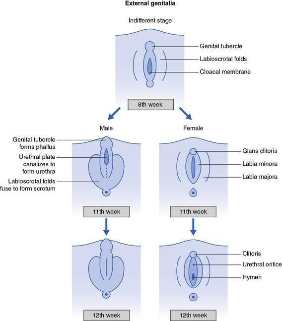
An understanding of normal sexual differentiation is essential in the evaluation of the child with a disorder of sexual development. Normal sexual development in the embryo consists of three related sequential processes:


Internal genitalia


Up until about 7 weeks, male and female embryos develop in an identical fashion, with bipotential gonads and both wolffian and müllerian internal genital ducts present (Fig. 19.3.1).
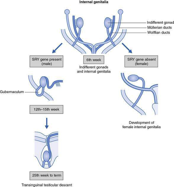

In males, the presence of the sex-determining region on the Y chromosome (SRY gene) directs the bipotential gonad to become a testis. At least four other genes are also required for normal testicular development. By 7–8 weeks the testis has recognizable tubules and starts producing androgens, including testosterone from the Leydig cells and müllerian-inhibiting substance (MIS) from the Sertoli cells. Circulating hormone levels are low and masculinization of the internal genital ducts occurs by locally acting (exocrine) secretion of these hormones down the wolffian duct.


High levels of testosterone promote the ipsilateral development of the wolffian duct to become the epididymis, vas deferens and seminal vesicle. High levels of MIS lead to ipsilateral müllerian duct regression. This process occurs during a critical period between 8 and 12 weeks. The Leydig cells also produce a relaxin-like factor that, together with MIS and androgens, masculinizes the gubernaculum. The gubernaculum holds the testis near the inguinal ligament during early development and from 25 weeks it begins to elongate, steering the testis towards the scrotum.


image Clinical example


A healthy, full-term infant was born with ambiguous genitalia. On examination there was an enlarged clitoris (2 cm in length), posterior fusion of the labia, with a single opening visible, and no gonads were palpable. The genitalia were noted to be hyperpigmented. There was no history of consanguinity but a previous male sibling had died at 2 weeks of age after a vomiting illness.


Initial investigations confirmed that the baby’s chromosomes were 46,XX and pelvic ultrasonography showed the presence of a normal uterus and ovaries. The 17-hydroxyprogesterone level at 48 hours of age was markedly increased, confirming the diagnosis of congenital adrenal hyperplasia due to 21-hydroxylase deficiency. Treatment with hydrocortisone was commenced. Daily electrolytes were normal until the 6th day of life, when hyperkalaemia developed, confirming the salt-wasting form of the condition. Fludrocortisone therapy was then added. The child was referred to a paediatric surgeon for consideration of corrective genital surgery. She will require lifelong replacement therapy and monitoring.


The hyperpigmentation was due to the adrenocorticotrophic hormone (ACTH) excess and resolved with adequate replacement therapy. It was likely that the previous male sibling had also had the condition, as it is inherited as an autosomal recessive trait. The genitalia of affected males are usually normal at birth apart from some hyperpigmentation, and affected male infants typically present with a salt-wasting crisis in the first few weeks of life. It is often confused with pyloric stenosis, which also presents at this age with vomiting and dehydration.


In females, the absence of testosterone and MIS leads to wolffian duct regression and müllerian duct preservation. The müllerian ducts develop into the uterus, fallopian tubes and upper vagina.


Aug 4, 2016 | Posted by in PEDIATRICS | Comments Off on The child of uncertain sex

Full access? Get Clinical Tree

Get Clinical Tree app for offline access