15.1 Suspected heart disease
assessment
Scale of the problem
Cardiac abnormalities or disease affect approximately 1% of children in the developed world and 2–3% in developing countries. This difference is largely due to the higher incidence of rheumatic heart disease in the latter.
The prevalence of congenital heart disease is approximately 8 in 1000 newborn infants. In Australia, heart disease remains one of the major causes of infant mortality.
Presentation
Significant heart disease in children presents with symptoms and signs of heart failure or cyanosis. These will, of course, vary depending on the age of the patient and severity of disease. Milder forms of heart disease may present by the detection of an asymptomatic murmur.
Assessment
History
It is important to determine the onset and type of symptoms. In babies, breathlessness, feeding difficulties, inability to complete feeds and poor weight gain are important to elucidate and may be indicative of a significant heart problem. Cyanosis due to heart disease is usually persistent but may intermittently increase in severity. Intermittent peripheral and circumoral cyanosis are common in normal children, typically occurring when cold (e.g. swimming) or conversely when febrile. Cyanosis may also be associated with breath-holding in children with normal hearts, and in these situations clearly defining the sequence of events leading up to the cyanotic episode is important.
Chest pain in children is rarely due to heart disease. The history of the chest pain is useful in distinguishing cardiac from non-cardiac chest pain; however, this may be difficult to elicit in young children. Pain consistently associated with exertion is more likely to be cardiac in nature, although its location may indicate a musculoskeletal rather than cardiac origin. Oesophageal and gastric pain may be indicated by the history. Brief episodes of chest discomfort may be associated with viral illnesses and myopericarditis.
Palpitations associated with collapse are clearly concerning but the majority of children presenting with palpitations do not lose consciousness with episodes of tachycardia. It is useful to determine the rate, duration, nature of onset and offset of these episodes, in addition to the circumstances surrounding them. Most children may be able to tap out with their hand how fast their heart rate is, or parents can be taught to measure the pulse and keep a diary of events.
Pulses
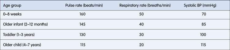
Examination of pulses should include both left and right arms and femoral pulses. The upper and lower limb pulses are best compared when palpated simultaneously. Relatively reduced lower-limb pulses suggest coarctation, but femoral pulses may be difficult to feel in the first few days of life. Bounding pulses due to a wide pulse pressure may be associated with patent ductus arteriosus, significant aortic regurgitation, and high cardiac output states. The physiological increase in heart rate in children varies markedly with activity and so it is the resting rate that should be noted (Table 15.1.1).
Blood pressure
Measurement of blood pressure should be a routine part of examination in children. Use of an appropriate cuff is vital. The balloon/bladder of the cuff should be wide enough to cover two-thirds of the upper arm. In practice, the largest cuff that can be fitted to the upper arm without covering the antecubital fossa is used. Blood pressure should normally be recorded by auscultation of Korotkoff sounds, as in adults, although palpation (of the brachial or radial pulse) may be employed to assess systolic pressure in young children and infants if auscultation proves to be difficult. Significant errors in blood pressure are more likely to result from the use of a cuff that is too small than one that is overlarge. Newer automated devices are able to detect the arterial pulsation and limit the upper pressure of inflation; however, these devices are still relatively slow and may overestimate blood pressure in restless or uncooperative children.
For measurement of leg blood pressure, a cuff may be placed on the thigh. An adult arm cuff may be large enough for young children, but larger children or adolescents will require a ‘thigh cuff’, which is larger than an adult arm cuff.
Normal blood pressure varies at different ages (see Table 15.1.1).
Palpation of the cardiac impulse
Location of the apex beat and documentation of any abnormal/forceful impulses/thrill of the precordium is important.
Auscultatory findings
Splitting of the second heart sound should be noted (Fig. 15.1.1). Splitting is widened during inspiration. Fixed splitting, a feature of atrial septal defect, implies absence of variation between inspiration and expiration (Fig. 15.1.2) and is also typically widely split.

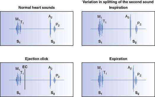
Fig. 15.1.1 Illustration of normal heart sounds, normal splitting of the second sound and ejection click (EC).

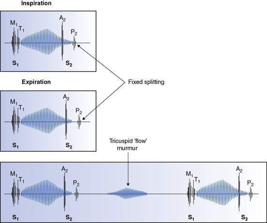
Fig. 15.1.2 Auscultatory signs associated with an atrial septal defect showing ejection systolic murmur, fixed splitting of S2 and tricuspid flow murmur (see Chapter 15.2).
Accentuation of the pulmonary component of the second sound tends to be associated with a loud second sound, which may be palpable, often with no definite splitting, and implies the presence of pulmonary hypertension. However, it should be noted that the normal aortic closure sound may be loud in children with a thin chest wall and is sometimes palpable at the upper left sternal border. The presence of an ejection click (see Fig. 15.1.1) is a useful ancillary auscultatory finding. Such sounds are heard shortly after the first heart sound and tend to be high-frequency and discrete in character. If heard at the apex, it usually implies a bicuspid aortic valve or aortic valve stenosis. When originating from the pulmonary valve, it is heard at the left sternal edge and varies with respiration, being louder on expiration. This finding is characteristic of pulmonary valve stenosis.
Murmurs
The following features of the murmur should be determined:
Timing
Murmurs may be systolic (limited to systole), diastolic (limited to diastole) or continuous (extending from systole into diastole). Note that continuous murmurs are not necessarily present throughout the cardiac cycle. Murmurs may be timed by simultaneously palpating a peripheral pulse.
Amplitude
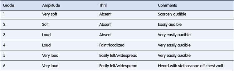
Murmurs may be graded according to the scale in Table 15.1.2. The amplitude of the murmur is affected by the thickness of the chest wall, and the direction, volume and velocity of blood flow relative to the stethoscope position.
Characterization
Ejection murmurs (Fig. 15.1.3) are systolic and crescendo–decrescendo in character, starting shortly after the first sound. Good examples are the murmurs of pulmonary or aortic valve stenosis.
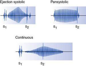
Pansystolic murmurs (Fig. 15.1.3) are murmurs that commence at the first sound and continue to the second sound. They may be due to atrioventricular valve incompetence (e.g. mitral incompetence) or a ventricular septal defect (VSD).
Diastolic murmurs may be early diastolic (see Fig. 15.1.4) (commencing at the second sound) or mid-diastolic (see Fig. 15.1.2). The former reflect either aortic or pulmonary incompetence, whereas mid-diastolic murmurs occur during ventricular filling and reflect either stenosis of or increased blood flow through an atrioventricular valve (e.g. mitral stenosis or secondary to a large left–right shunt due to a VSD).

Stay updated, free articles. Join our Telegram channel

Full access? Get Clinical Tree

