11.5 Surgical conditions in the newborn
The majority of the conditions discussed in this chapter will present initially to the paediatrician, general practitioner or obstetrician as emergencies. Delay in diagnosis may seriously compromise recovery and will almost certainly increase morbidity. Disorders that are obvious at birth but do not require urgent surgical referral have not been included in this chapter. For information on these, the reader is referred to paediatric surgical texts.
Oesophageal atresia
Any newborn infant who appears to salivate excessively (drooling) at birth should be suspected of having oesophageal atresia. This is a congenital abnormality where the mid-portion of the oesophagus is missing. In most there is an abnormal communication between the trachea and the lower oesophageal segment, called a distal tracheo-oesophageal fistula.
The diagnosis is confirmed by passing a large, firm catheter, for example a 10-French gauge orogastric tube, through the mouth and finding that it cannot be passed more than about 10 cm from the gums. The child must not be fed; otherwise, aspiration of feeds into the lungs is likely to occur. A plain X-ray of the torso will show gas in the bowel, confirming the presence of a distal tracheo-oesophageal fistula. About 50% of these infants have other congenital abnormalities, most of which form part of the VACTERL association (vertebral, anorectal, cardiac, renal and limb abnormalities; see Chapter 10.3). Major chromosomal abnormalities are seen in 5%, of which trisomy 18 and trisomy 21 are the most frequent. Many are premature and a history of maternal polyhydramnios is common.
Duodenal obstruction
Bile-stained vomiting starts soon after birth. The obstruction may be:
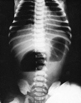
In duodenal atresia there may be other abnormalities, such as Down syndrome and imperforate anus (see Chapter 10.3). In the absence of birth asphyxia these infants are usually alert and feed well, but they vomit bile-stained material almost immediately. There may be epigastric distension. The diagnosis of duodenal atresia is made on plain X-ray of the abdomen, which reveals a characteristic ‘double bubble’ due to gas in the stomach and proximal duodenum (Fig. 11.5.1). Little or no gas will be visible distal to the obstruction. Duodenoduodenostomy is performed after resuscitation and correction of any fluid or electrolyte disturbance.

Fig. 11.5.1 X-ray of the abdomen in a neonate demonstrating the ‘double bubble’ sign. Note the absence of gas in the bowel distal to the second bubble. This child had duodenal atresia.
Bile-stained vomiting may also be an indication of malrotation complicated by volvulus. The small bowel mesentery has a narrow attachment to the posterior abdominal wall, the so-called ‘universal mesentery’, which allows the midgut to twist around the superior mesenteric vessels. This is a true surgical emergency as the blood supply to the midgut may be cut off as the midgut twists around this axis. The diagnosis can be confirmed with an urgent barium meal or by ultrasonography. If signs of peritonitis with abdominal distension and guarding are already present, the infant should be taken immediately to theatre.
Distal bowel obstruction
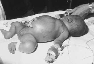
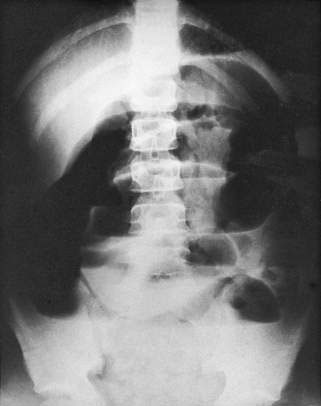
In more distal bowel obstructions, vomiting remains a major feature but tends to occur later and is associated with abdominal distension. The more distal the obstruction, the later the vomiting and the more pronounced the distension (Fig. 11.5.2). The vomitus may become faeculent. An erect film of the abdomen will show distended loops of bowel and fluid levels (Fig. 11.5.3). The number of loops is dependent on the level of obstruction. The radiological appearances of Hirschsprung disease, meconium ileus and ileal atresia may be similar, and a contrast study, rectal biopsy or laparotomy may be required to make the definitive diagnosis.

Fig. 11.5.2 Distal bowel obstruction in a neonate. Gross abdominal distension was evident and associated with vomiting of faeculent material.

Fig. 11.5.3 Erect X-ray of the abdomen, showing marked dilatation of multiple loops of bowel and several fluid levels. The most likely diagnoses include Hirschsprung disease, meconium ileus and ileal atresia.
Hirschsprung disease
Hirschsprung disease (congenital megacolon) is the most common cause of neonatal bowel obstruction. There is an absence of ganglion cells for a variable distance proximal to the anus. Peristalsis is abnormal in the aganglionic segment and results in an incomplete lower intestinal obstruction with severe constipation. The bowel proximal to the aganglionic segment becomes dilated and hypertrophied. The diagnosis is confirmed on suction rectal biopsy. Most of these infants present at 3 or 4 days of age with increasing abdominal distension and delay in the passage of meconium. Surgical correction involves:
It is often performed as a single-stage procedure at diagnosis in the neonate, but in certain circumstances requires a staged approach.

Stay updated, free articles. Join our Telegram channel

Full access? Get Clinical Tree

