Fig. 16.1
Pelvic lymph node dissection. 1. Sigmoid-descending colon. 2. Common iliac nodes. 3. External iliac artery. 4. External iliac vein. 5. Obturator nodes. 6. Internal iliac artery
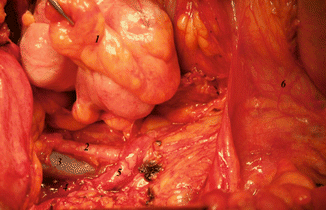
The bowel is packed to the opposite side of dissection. The colon and ureter are retracted with a large Deaver retractor toward the opposite shoulder. The retroperitoneum is entered by retracting the round ligament and incising the peritoneum lateral to the ovarian vessels up to the pelvic brim. The loose areolar tissue is dissected to identify the pelvic vessels and ureter. The paravesical space is identified by dissecting down into the obturator fossa retracting the obliterated umbilical artery and lateral part of the bladder medially and external iliac vessels laterally. The pararectal space is opened by bluntly dissecting between the ureter and internal iliac artery.
The extent of lymph node dissection is genitofemoral nerve laterally, muscular pelvic wall posterolaterally, obturator nerve posteriorly, and circumflex iliac vein crossing the external iliac artery or the level where the external iliac vessels exit the abdomen distally. Midpoint of the common iliac artery serves as a divide between pelvic and para-aortic nodes. The lymph nodes are dissected using a right angled clamp and feeding vessels cauterized using bipolar cautery and excised. This includes the tissues anterior and medial to the common iliac artery, anterior and medial to external iliac artery, medial to external iliac vein, and anterior to the internal iliac artery extending distally along the superior vesical artery (Fig. 16.2). The external iliac vein is retracted anteriorly with a vein retractor. Blunt dissection is performed within the obturator space to reveal the course of the obturator nerve. The lymph nodes anterior to the obturator nerve are removed.


Fig. 16.2
Completed pelvic node dissection. 1. Cecum. 2. External iliac artery. 3. External iliac vein. 4. Internal iliac vein. 5. Ureter. 6. Beginning of jejunum
Para-aortic Lymph Node Dissection
The extent of para-aortic lymph node dissection is from the aortic bifurcation up to either the inferior mesenteric artery or preferably to the level of renal veins.
The small bowel loops are brought out of the incision, packed inside a one meter sheet, and retracted into the upper abdomen using a Deaver retractor. The peritoneal incision is extended along the common iliac vessels up the aorta to the root of the small bowel and on the right side along the paracolic gutter. Midline incision may also be used for aortocaval dissection up to the level of origin of right ovarian vessels. The small bowel loops and right colon are then moved out of the field and right ovarian vessels and ureter freed from the posterior aspect of right colon. On the left side, exposure is attained by extending peritoneal incision through left paracolic gutter and retracting the left colon laterally. The dissection is started from the aortic bifurcation and proceeded in a caudal to cephalad direction.
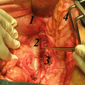
The lymphatic tissue from anterior surface of vena cava can be removed using sharp and blunt dissection. Care should be taken to prevent tearing of small venous branches entering the vena cava. There is a fairly constant vein within the lymphatics just above the bifurcation named the “Fellow’s vein,” which when torn can cause unexpectedly large defects in vena cava resulting in heavy bleeding. The inferior mesenteric artery is preserved, and dissection should be continued preferably up to the level of renal veins (Fig. 16.3).


Fig. 16.3
Para-aortic node dissection: 1.duodenum 2.inferior venacava 3. aorta 4. sigmoid & descending colon mesentry
Ligation of lumbar arteries above renal vessels should be avoided to prevent spinal cord ischemia. The intraoperative complications of retroperitoneal lymph node dissection are injury to ureter, pelvic vessels, obturator nerve, lumbar vein, aorta, and vena cava. Laceration of small veins (Fellow’s vein) is the common IVC injury. In case of vascular injury, the site of injury is compressed while sutures are ready. The area above and below is cleared, and the injured area is sutured with 5–0 or 6–0 PROLENE. For aortic rents, it is wise to get the help of a vascular surgeon. The postoperative complications include venous thrombosis, lymphocyst formation, and small bowel obstruction.
Special Considerations
1.


Uterine papillary serous carcinomas behave in a manner similar to ovarian cancers demonstrating spread within the peritoneal cavity even when the primary is confined to the endometrium. Hence, staging of uterine papillary serous carcinoma should include hysterectomy with bilateral salpingo-oophorectomy, peritoneal washings and biopsies, bilateral pelvic and para-aortic lymph node dissection, and omentectomy as is typically performed for ovarian cancer. In early-stage endometrial cancer, the majority of omental metastases consist of microscopic disease. Factors significantly associated with omental metastasis were adnexal spread, cul-de-sac implantation, papillary serous carcinoma, positive retroperitoneal lymph nodes, and grade 3 tumor. For patients with high-risk variables, a complete omentectomy should be considered [2].
Stay updated, free articles. Join our Telegram channel

Full access? Get Clinical Tree


