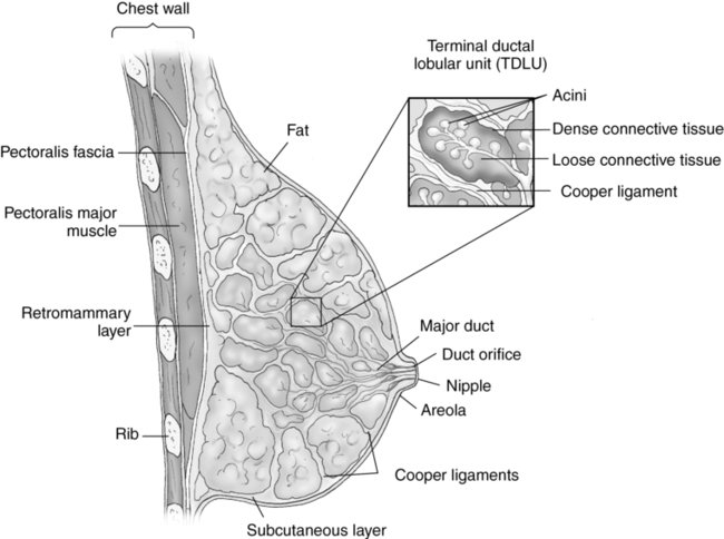
CHAPTER 15 protrusion of peritoneal contents through a defect in the abdominal wall. attaches the gastrocnemius and soleus muscles. smallest functional unit of the breast. a synovial cyst adjacent and posterior to the knee joint. Development Displacement of the Hip (DDH) a small filamentous fiber that is often a component of a cell. the presence of a single or multiple palpable cysts in the breast. a cyst caused by obstruction of a lactating duct. small tumor or fluid collection that can occur at the connection of any tendon an abnormal enlargement of a male breast or breasts. one of many channels that carry milk for the lobes of each breast to the nipple. an area of enlargement in a lactiferous duct near the areola. a flexible band of fibrous tissue binding joints together; provides flexibility to a joint. a collection of lobules within the breast parenchyma; approximately 15 to 20 lobes per breast. the simplest functional unit of the breast. breast parenchyma lying within the superficial fascia. a nonneoplastic fusiform enlargement of a digital branch of the medial or lateral plantar nerves. one of a pair of anterolateral abdominal wall muscles located lateral to the linea alba. located between the posterior margin of the mammary zone and the pectoralis muscles. a painful wrenching or laceration of the ligaments of a joint. to injure or impair by overuse or overexertion; wrench. double-walled tubular structures surrounding some tendons. bands of dense, fibrous connective tissue that attach muscle to bone. terminal ductal lobular unit (TDLU) small lobular unit formed by the acini and the terminal ducts. • Skin line appears hyperechoic. • Superficial and deep fascial planes appear hyperechoic. • Glandular breast parenchyma appears moderately hyperechoic. • Fat breast lobules should demonstrate a medium-gray echo pattern. • Retromammary layer appears hypoechoic. • Pectoralis muscles appear moderately hyperechoic. • Cooper ligaments appear as hyperechoic linear structures; may demonstrate posterior acoustic shadowing. • Lactiferous ducts appear as nonvascular, anechoic tubular structures coursing toward the nipple. • 7.5-MHz or higher linear transducer to obtain optimal resolution for penetration depth. • Proper image depth with focal zone(s) at or below the place of interest. • Gain settings demonstrating breast fat as a medium shade of gray and a simple cyst as an anechoic mass. • Increase in dynamic range setting. • Sufficient imaging depth to visualize structures immediately posterior to the region of interest. • Harmonic imaging can be used to reduce artifactual echoes within anechoic structures. • Spatial compounding can be used to improve visualization of structures posterior to a highly attenuating structure. • Proper Doppler controls for low-flow velocity (pulse repetition frequency [PRF], gain, wall filters). • Patient is generally placed in a right or left posterior oblique position. • Standoff pad should not exceed 1.0 cm in thickness. • Evaluation and documentation of breast parenchyma in two imaging planes, remaining perpendicular to the chest wall. • Proper annotation of the image location and scanning plane. • Images are generally labeled by quadrant and/or the face of a clock. • Distance from the nipple is described as 1, 2, or 3 (1 is closest to nipple). • Depth of the area of interest is described as A, B, or C (C is closest to the chest wall). • Documentation and measurement of any abnormality in two scanning planes should be included. • Color Doppler imaging to evaluate abnormalities for internal and peripheral flow. • Evaluate mass from a previous medical imaging study (i.e., mammogram). • Ultrasound-guided interventional procedure. • Evaluate male breast parenchyma.
Superficial structures: Breast, abdominal wall, and musculoskeletal sonography
The breast
Sonographic appearance of the breast
Technique
Examination technique and image optimization
Indications for a breast examination
PATHOLOGY FINDINGS
ETIOLOGY
CLINICAL FINDINGS
SONOGRAPHIC FINDINGS
DIFFERENTIAL CONSIDERATIONS
Cyst
Obstruction of a duct
Infection
Common around 35-50 yrs of age
Asymptomatic
Breast pain or tenderness
Palpable mass
Anechoic round or oval mass
Smooth, thin wall margins
Posterior acoustic enhancement
No internal vascular flow
Compresses with transducer pressure
Mass does not breach fascial plane(s)
May demonstrate internal echoes
Lactiferous duct
Fibroadenoma
Carcinoma
Cystosarcoma phyllodes
Uncommon benign fibroepithelial neoplasm
May undergo malignant transformation
Sudden onset of a palpable nontender breast mass
Mobile mass
Oval mass demonstrating a low- to medium-level echo pattern
Unilateral mass
May demonstrate cystic spaces within the mass
Smooth wall margins
Width of mass is larger than the height
Mass does not breach fascial plane(s)
Internal blood flow may be demonstrated
Complex cyst
Fibroadenoma
Carcinoma
Normal breast fat
Fibroadenoma
Tumor composed of dense epithelial and fibroblastic tissue
Influenced by estrogen levels
Asymptomatic
Palpable nontender breast mass
Mobile mass
Firm or rubbery on palpation
Solid oval-shaped breast mass
Low- to medium-level echo pattern
Posterior acoustic enhancement
Mass does not breach the fascial plane
Width of mass is larger than the height
Can degenerate or calcify
Internal blood flow may be demonstrated
Complex cyst
Normal breast fat
Carcinoma
Fibrocystic disease
Presence of palpable breast cyst(s)
Not generally associated with future development of breast carcinoma
Painful or tender breasts frequently 7-10 days before the start of menses
Increase in pain intensity closer to the start of menses
Hyperechoic breast parenchyma
Dense breast tissue
Prominent ducts
Numerous breast cysts
Multiple breast cysts
Mastitis
Hamartoma
Proliferation of normal tissues
Asymptomatic
Palpable mass
Heterogeneous complex mass
Smooth wall margins
May demonstrate posterior acoustic shadowing
Mass does not breach fascial plane(s)
Mass compresses with moderate transducer pressure
Complex cyst
Carcinoma
Fibroadenoma
Galactocele
Obstruction of a lactating duct
Palpable retroareolar mass
Round or oval hypoechoic retroareolar mass
Smooth wall margins
Posterior acoustic enhancement
Fibroadenoma
Complex cyst
Abscess
Gynecomastia
Abnormal proliferation of ductal, glandular tissue, and stroma
Increased amount of subcutaneous fat
Hormone disorders
Endocrine disorders
Neoplasms
Abnormal enlargement of the male breast(s)
Painful or tender breast(s)
Hypoechoic to hyperechoic tissue beneath the areola
Ducts converging toward the areola
Increased amount of breast fat
Unilateral or bilateral
Neoplasm
Mastitis
Lipoma
Mature adipose tissue
Soft, mobile mass
Homogeneous hyperechoic mass within the subcutaneous fat
Oval in shape
Smooth wall margins
May appear similar to breast fat
Glandular breast tissue
Fibroadenoma
Complex cyst
Mastitis
![]()
Stay updated, free articles. Join our Telegram channel

Full access? Get Clinical Tree

 Get Clinical Tree app for offline access