Chapter 174 Staphylococcus
Staphylococci are hardy, aerobic, gram-positive bacteria that grow in pairs and clusters and are ubiquitous as normal flora of humans and present on fomites and in dust. They are resistant to heat and drying and may be recovered from nonbiologic environments weeks to months after contamination. Strains are classified as Staphylococcus aureus if they are coagulase positive or as 1 of the many species of coagulase-negative staphylococci (e.g., Staphylococcus epidermidis, Staphylococcus saprophyticus, Staphylococcus haemolyticus, etc). Often, S. aureus produces a yellow or orange pigment and β-hemolysis on blood agar and S. epidermidis produces a white pigment with variable hemolysis results, although definitive species confirmation requires further testing. S. aureus has many virulence factors that mediate various serious diseases, whereas coagulase-negative staphylococci tend to be less pathogenic unless an indwelling foreign body (e.g., intravascular catheter) is present. Emerging antimicrobial resistance has become important, especially to the β-lactam antibiotics and less often to vancomycin.
174.1 Staphylococcus aureus
S. aureus is the most common cause of pyogenic infection of the skin and soft tissue, causing impetigo, furuncles (boils), cellulitis, abscess, lymphadenitis, paronychia, omphalitis, and wound infection. Bacteremia (primary and secondary) is common and can be associated with or result in osteomyelitis, suppurative arthritis, deep abscesses, pneumonia, empyema, endocarditis, pyomyositis, pericarditis, and rarely meningitis. Toxin-mediated diseases, including food poisoning, staphylococcal scarlet fever, scalded skin syndrome, and toxic shock syndrome (TSS), are caused by certain S. aureus strains. Methicillin resistance is a global problem.
Etiology
Disease may result from tissue invasion or injury caused by various toxins and enzymes produced by the organism. Strains of S. aureus can be identified by the virulence factors they produce and can be classified by various molecular techniques.
Adhesion of S. aureus to mucosal cells is mediated by teichoic acid in the cell wall; exposure to the submucosa or subcutaneous sites increases adhesion to fibrinogen, fibronectin, collagen, and other proteins. Different strains of S. aureus produce many different virulence factors that have 1 or more of 4 different roles: protect the organism from host defenses, localize infection, cause local tissue damage, and act as toxins affecting noninfected tissue sites.
Most strains of S. aureus possess factors that protect the organism from host defenses. Many staphylococci produce a loose polysaccharide capsule, or slime layer, which may interfere with opsonophagocytosis. Production of coagulase and/or clumping factor differentiates S. aureus from S. epidermidis and other coagulase-negative staphylococci. Clumping factor interacts with fibrinogen to cause large clumps of organisms, interfering with effective phagocytosis. Coagulase causes plasma to clot by interacting with fibrinogen and this may have an important role in localization of infection (abscess formation). Protein A is present in most strains of S. aureus but not coagulase-negative staphylococci and reacts specifically with immunoglobulin G1 (IgG1), IgG2, and IgG4. It is located on the outermost coat of the cell wall and can absorb serum immunoglobulins, preventing antibacterial antibodies from acting as opsonins and thus inhibiting phagocytosis. Other enzymes elaborated by staphylococci include catalase (inactivates hydrogen peroxide, promoting intracellular survival), penicillinase or β-lactamase (inactivates penicillin at the molecular level), and lipase (associated with skin infection).
Many strains of S. aureus produce substances that cause local tissue destruction. A number of immunologically distinct hemolysins that act on cell membranes and cause tissue necrosis have been identified (α-toxin, β-hemolysin, δ-hemolysin). Panton-Valentine leukocidin (PVL), which is produced by many current strains of S. aureus and has been associated with invasive skin disease, combines with the phospholipid of the phagocytic cell membrane, producing increased permeability, leakage of protein, and eventual death of the cell.
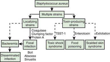
Many strains of S. aureus release 1 or more exotoxins. Exfoliatins A and B are serologically distinct proteins that produce localized (bullous impetigo) or generalized (scalded skin syndrome, staphylococcal scarlet fever) dermatologic complications (Chapter 651). Exfoliatins produce skin separation by splitting the desmosome and altering the intracellular matrix in the stratum granulosum.
Toxic shock syndrome toxin-1 (TSST-1) is associated with TSS related to menstruation and focal staphylococcal infection. TSST-1 is a superantigen that induces production of interleukin-1 and tumor necrosis factor, resulting in hypotension, fever, and multisystem involvement. Enterotoxin A and enterotoxin B also may be associated with nonmenstrual TSS.
Epidemiology
Many neonates are colonized within the 1st wk of life, and 20-40% of normal individuals carry at least 1 strain of S. aureus in the anterior nares at any given time.
The organisms may be transmitted from the nose to the skin, where colonization seems to be more transient. Persistent umbilical, vaginal, and perianal carriage may occur.
Heavily colonized nasal carriers (often aggravated by a viral upper respiratory tract infection) are particularly effective disseminators. Exposure to S. aureus generally occurs by auto-inoculation or direct contact with the hands of other colonized individuals. Handwashing between patient contacts is essential to decrease the nosocomial spread of staphylococci. Spread via fomites is rare.
Invasive disease may follow colonization. Antibiotic therapy with a drug to which S. aureus is resistant favors colonization and the development of infection. Other factors that increase the likelihood of infection include wounds, skin disease, ventriculoperitoneal shunts, intravenous or intrathecal catheterization, corticosteroid treatment, malnutrition, and azotemia. Viral infections of the respiratory tract, especially influenza virus, may predispose to secondary bacterial infection with staphylococci.
Pathogenesis
The development of staphylococcal disease is related to resistance of the host to infection and to virulence of the organism (Fig. 174-1). The intact skin and mucous membranes serve as barriers to invasion by staphylococci. Defects in the mucocutaneous barriers produced by trauma, surgery, foreign surfaces (sutures, shunts, intravascular catheters), and burns increase the risk for infection.

Figure 174-1 Relationship of virulence factors and diseases associated with Staphylococcus aureus. TSST-1, toxic shock syndrome toxin-1.
Infants may acquire type-specific humoral immunity to staphylococci transplacentally. Older children and adults develop antibodies to staphylococci as a result of colonization or minor infections. Antibody to the various S. aureus toxins appears to protect against those specific toxin-mediated diseases, but humoral immunity does not necessarily protect against focal or disseminated S. aureus infection with the same organisms.
Congenital defects in chemotaxis (Job syndrome, Chédiak-Higashi syndrome, Wiskott-Aldrich syndrome) and defective phagocytosis and killing (neutropenia, chronic granulomatous disease) increase the risk for staphylococcal infections. Patients with HIV infection have neutrophils that are defective in their ability to kill S. aureus in vitro. Patients with recurrent staphylococcal infection should be evaluated for immune defects, especially those involving neutrophil dysfunction.
Clinical Manifestations
The signs and symptoms vary with the location of the infection, which is most commonly the skin but may be any tissue. Disease states of various degrees of severity are generally a result of local suppuration, systemic dissemination with metastatic infection, or systemic effects of toxin production. Although the nasopharynx and skin of many persons may be colonized with S. aureus, disease due to this organism is relatively uncommon. Skin infections due to S. aureus are considerably more prevalent among persons living in low socioeconomic circumstances and particularly among those in tropical climates.
Skin
S. aureus is an important cause of pyogenic skin infections, including impetigo contagiosa, ecthyma, bullous impetigo, folliculitis, hydradenitis, furuncles, carbuncles, staphylococcal scalded skin syndrome, and staphylococcal scarlet fever. Infection may also complicate wounds or occur as superinfection of other noninfectious skin disease (eczema). Recurrent furunculosis is associated with repeated episodes of pyoderma over months to years. Recurrent skin and soft tissue infections are commonly noted with community-associated methicillin-resistant S. aureus (MRSA) and often affect the lower extremities and buttocks. S. aureus is also an important cause of wound infections and can cause deep soft tissue involvement, including cellulitis and rarely necrotizing fasciitis.
Respiratory Tract
Infections of the upper respiratory tract due to S. aureus are rare, in particular considering the frequency with which the anterior nares are colonized. In normal hosts, otitis media (Chapter 632) and sinusitis (Chapter 372) are rarely caused by S. aureus. S. aureus sinusitis is relatively common in children with cystic fibrosis or defects in leukocyte function and may be the only focus of infection in some children with toxic shock syndrome. Suppurative parotitis is a rare infection, but S. aureus is a common cause. A membranous tracheitis that complicates viral croup may be due to infection with S. aureus, but other organisms are also possible. Patients typically have high fever, leukocytosis, and evidence of severe upper airway obstruction. Direct laryngoscopy or bronchoscopy shows a normal epiglottis with subglottic narrowing and thick, purulent secretions within the trachea. Treatment requires careful airway management and appropriate antibiotic therapy.
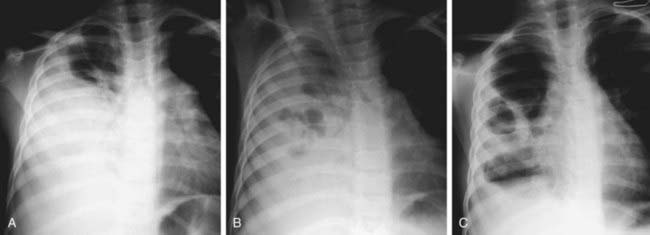
Pneumonia (Chapter 392) due to S. aureus may be primary (hematogenous) or secondary after a viral infection such as influenza. Hematogenous pneumonia may be secondary to septic emboli from right-sided endocarditis or septic thrombophlebitis, with or without the presence of intravascular devices. Inhalation pneumonia is caused by alteration of mucociliary clearance (see cystic fibrosis, Chapter 395), leukocyte dysfunction, or bacterial adherence initiated by a viral infection. Common symptoms and signs include high fever, abdominal pain, tachypnea, dyspnea, and localized or diffuse bronchopneumonia or lobar disease. S. aureus often causes a necrotizing pneumonitis that may be associated with development of empyema, pneumatoceles, pyopneumothorax, and bronchopleural fistulas.
Sepsis
S. aureus bacteremia and sepsis may be primary or associated with any localized infection. The onset may be acute and marked by nausea, vomiting, myalgia, fever, and chills. Organisms may localize subsequently at any site (usually a single deep focus) but are found especially in the heart valves, lungs, joints, bones, and abscesses.
In some instances, especially in young adolescent males, disseminated S. aureus disease occurs, characterized by fever, persistent bacteremia despite antibiotics, and focal involvement of 2 or more separate tissue sites (skin, bone, joint, kidney, lung, liver, heart).
In these cases, endocarditis and septic thrombophlebitis must be ruled out.
Muscle
Localized staphylococcal abscesses in muscle associated with elevation of muscle enzymes sometimes without septicemia have been called pyomyositis. This disorder has been reported most frequently from tropical areas but also occurs in the USA in otherwise healthy children. Multiple abscesses occur in 30-40% of cases. History may include prior trauma at the site of the abscess. Surgical drainage and appropriate antibiotic therapy are essential.
Bones and Joints
S. aureus is the most common cause of osteomyelitis and suppurative arthritis in children (Chapters 676 and 677).
Central Nervous System
Meningitis (Chapter 595.1) caused by S. aureus is not common; it is associated with penetrating cranial trauma and neurosurgical procedures (craniotomy, cerebrospinal fluid [CSF] shunt placement) and less frequently with endocarditis, parameningeal foci (epidural or brain abscess), diabetes mellitus, or malignancy. The CSF profile of S. aureus meningitis is indistinguishable from that in other forms of bacterial meningitis.
Heart
S. aureus is a common cause of acute endocarditis (Chapter 431) on native valves. Perforation of heart valves, myocardial abscesses, heart failure, conduction disturbances, acute hemopericardium, purulent pericarditis, and sudden death may ensue.
Kidney
S. aureus is a common cause of renal and perinephric abscess (Chapter 532), usually of hematogenous origin. Pyelonephritis and cystitis due to S. aureus are unusual.
Toxic Shock Syndrome (TSS)
S. aureus is the principal cause of TSS (Chapter 174.2), which should be suspected in anyone with fever, shock, and/or a scarlet fever-like rash.
Intestinal Tract
Staphylococcal enterocolitis rarely follows overgrowth of normal bowel flora by S. aureus, which can occur as a result of broad-spectrum oral antibiotic therapy. Diarrhea is associated with blood and mucus. Peritonitis associated with S. aureus in patients receiving long-term ambulatory peritoneal dialysis usually involves the catheter tunnel. Removal of the catheter is required to achieve a bacteriologic cure.
Food poisoning (Chapter 332) may be caused by ingestion of preformed enterotoxins produced by staphylococci in contaminated foods. Approximately 2-7 hr after ingestion of the toxin, sudden, severe vomiting begins. Watery diarrhea may develop, but fever is absent or low. Symptoms rarely persist longer than 12-24 hr. Rarely, shock and death may occur.
Diagnosis
The diagnosis of S. aureus infection depends on isolation of the organism from nonpermissive sites such as cellulitis aspirates, abscess cavities, blood, bone or joint aspirates, or other sites of infection. Swab cultures of surfaces are not as useful, as they may reflect surface contamination rather than the true cause of infection. Tissue samples or fluid aspirates in a syringe provide the best culture material. Isolation from the nose or skin does not necessarily imply causation because these sites may be normally colonized sites. Because of the high prevalence of MRSA, the increasing severity of S. aureus infections, and the fact that bacteremia is not universally present even in severe S. aureus infections, it is usually important to obtain a nonpermissive culture of any potential focus of infection as well as a blood culture prior to starting antibiotic treatment. The organism can be grown readily in liquid and on solid media. After isolation, identification is made on the basis of Gram stain and coagulase, clumping factor, and protein A reactivity. Patterns of susceptibility to antibiotics should be assessed in serious cases, as antimicrobial resistance is increasingly common.
Diagnosis of S. aureus food poisoning is made on the basis of epidemiologic and clinical findings. Food suspected of contamination should be cultured and can be tested for enterotoxin.
Differential Diagnosis
Skin lesions due to S. aureus may be indistinguishable from those due to group A streptococci; the former usually expand slowly, while the latter are more prone to spread rapidly. S. aureus pneumonia can be suspected on the basis of chest roentgenograms that reveal pneumatoceles, pyopneumothorax, or lung abscess (Fig. 174-2). Fluctuant skin and soft tissue lesions also can be caused by other organisms, including Mycobacterium tuberculosis, atypical mycobacteria, Bartonella henselae (cat-scratch disease), Francisella tularensis, and various fungi, among others.

Figure 174-2 Pneumatocele formation. A, A 5 yr old child with Staphylococcus aureus pneumonia initially demonstrated consolidation of the right middle and lower zones. B, Seven days later, multiple lucent areas are noted as pneumatoceles develop. C, Two wk later, significant resolution is evident, with a rather thick-walled pneumatocele persisting in the right midzone associated with significant residual pleural thickening.
(From Kuhn JP, Slovis TL, Haller JO: Caffey’s pediatric diagnostic imaging, vol 1, ed 10, Philadelphia, 2004, Mosby, pp 1003–1004.)

Full access? Get Clinical Tree

