Skin and Soft Tissue Infections
Marc H. Lebel
EPIDEMIOLOGY AND PATHOGENESIS
The most common manifestations of skin and soft tissue bacterial infections are presented in Box 74.1. Initial colonization of the skin occurs during delivery and reflects the organisms present in the birth canal. Subsequently, acquisition of Staphylococcus aureus and other bacteria occurs by contamination from the environment, the hands of the parents, and the personnel. The first site of colonization is the umbilical cord. In most infants, this is not followed by an infection.
BOX 74.1 Infections of the Skin and Soft Tissues
Pustular dermatitis and epidemic pustulosis
Bullous impetigo
Staphylococcal scalded skin syndrome (Ritter disease)
Staphylococcal pyoderma
Neonatal folliculitis
Scarlatiniform eruptions
Abscess of skin or scalp
Mastitis or breast abscess
Omphalitis
Cellulitis
Adenitis
Necrotizing fasciitis
Ecthyma gangrenosum
CLINICAL MANIFESTATIONS AND THERAPY
Impetigo
The most frequent skin and soft tissue infection is impetigo neonatorum (see Fig. 74.1). This presents with pustular lesions that cluster around the umbilicus and diaper area. It is not usually associated with fever or systemic illness. Risk factors include abrasions of the skin following forceps use, scalp wounds from intrapartum fetal monitoring devices, degeneration of the umbilical cord, intravascular catheter, and circumcision. Gram-positive and polymorphonuclear leukocytes present on Gram-stained smear examination distinguish pustules from erythema toxicum, in which case only eosinophils are seen on the smear. If only a few lesions are present, they are treated effectively by cleansing alone (chlorhexidine solution) or cleansing followed by application of topical antibiotic ointment. More extensive lesions may be treated with oral cephalexin for 5 to 10 days. Bullous impetigo is more likely to spread and should be treated with intravenous antibiotics in the absence of prompt response to local or oral therapy.
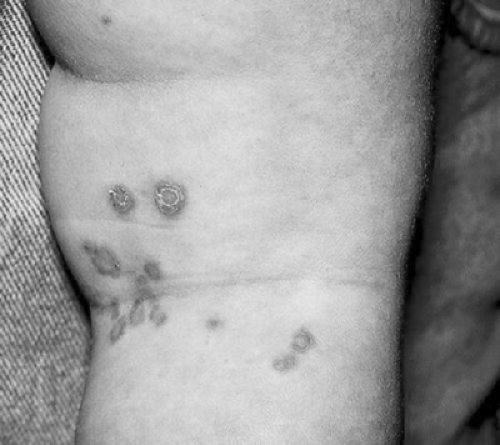 FIGURE 74.1. Impetigo. This photo does not show intact blisters, only the flaccid remains of the lesions. (From Goodheart HP. Goodheart’s photoguide of common skin disorders, 2nd ed. Philadelphia: Lippincott Williams & Wilkins, 2003.) |
Staphylococcal Scalded Skin Syndrome
Staphylococcal scalded skin syndrome, or Ritter disease, is a much more extensive, exfoliative disease caused by phage group II staphylococci (see also Chapter 68, Dematologic Diseases). These organisms elaborate and release a circulating exotoxin that leads to blistering of the upper layer of the skin (stratum granulosum). Infants first present with a tender, sunburn-like erythematous rash. Then bullae develop with desquamation of large areas. Lesions commonly affect the flexures; a large portion of the skin can be affected (Fig. 74.2). The presence of Nikolsky sign (desquamation of the superficial layer of skin after light pressure over areas that appear to be uninvolved) is pathognomonic. The site of toxin production is remote from the area of involvement and is usually the conjunctiva, nasopharynx, umbilicus, or circumcision site. Staphylococcal bacteremia and superinfection are absent. These infants require intravenous therapy to eradicate the source of the toxin.
Stay updated, free articles. Join our Telegram channel

Full access? Get Clinical Tree


