Chapter 411 Skeletal Diseases Influencing Pulmonary Function
Pulmonary function is influenced by the structure of the chest wall (Chapter 365). Chest wall abnormalities can lead to restrictive or obstructive pulmonary disease, impaired respiratory muscle strength, and decreased ventilatory performance in response to physical stress. The congenital chest wall deformities include pectus excavatum, pectus carinatum, sternal clefts, Poland syndrome, and skeletal and cartilage dysplasias. Vertebral anomalies such as kyphoscoliosis can alter pulmonary function in children and adolescents.
411.1 Pectus Excavatum (Funnel Chest)
Etiology
Midline narrowing of the thoracic cavity is usually an isolated skeletal abnormality. The cause is unknown. Pectus excavatum can occur in isolation or it may be associated with a connective tissue disorder (Marfan [Chapter 693] or Ehlers-Danlos syndrome [Chapter 651]). It may be acquired secondarily to chronic lung disease, neuromuscular disease, or trauma.
Clinical Manifestations
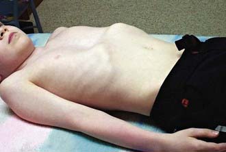
The deformity is present at or shortly after birth but is usually not associated with any symptoms at that time. In time, decreased exercise tolerance, fatigue, chest pain, palpitations, recurrent respiratory infections, wheezing, stridor, and cough may be present. Because of the cosmetic nature of this deformity, children may experience significant psychologic stress. Physical examination may reveal sternal depression, protracted shoulders, kyphoscoliosis, inferior rib flares, rib cage rigidity, forward head tilt, scapular winging, and loss of vertebral contours (Fig. 411-1). Patients exhibit paroxysmal sternal motion and a shift of point of maximal impulse to the left. Innocent systolic murmurs may be heard.
Laboratory Findings
Lateral chest radiograms demonstrate the sternal depression. Use of the Haller index on chest CT (maximal internal transverse diameter of the chest divided by the minimal anteroposterior diameter at the same level) in comparison with age- and gender-appropriate normative values for determining the extent of depression of the chest wall anomaly has become useful in determining the extent of the anatomic abnormality. An electrocardiogram may show a right-axis deviation or Wolff-Parkinson-White syndrome (Chapter 429); an echocardiogram may demonstrate mitral valve prolapse (Chapter 422.3) and ventricular compression. Results of static pulmonary function tests may be normal but commonly show an obstructive defect in the lower airways and, less commonly, a restrictive defect due to abnormal chest wall mechanics. Exercise testing may demonstrate either normal tolerance or limitations from underlying cardiopulmonary dysfunction that appear associated with the severity of the defect. Ventilatory limitations are commonly seen in younger children and adolescents, whereas cardiac limitations secondary to stroke volume impairments are more commonly seen in older adolescents and young adults.
Brigato RR, Campos JR, Jatene FB, et al. Pectus excavatum: evaluation of Nuss technique by objective methods. Interact Cardiovasc Thorac Surg. 2008;7:1084-1088.
Borowitz D, Cerny F, Zallen G, et al. Pulmonary function and exercise response in patients with pectus excavatum after Nuss repair. J Pediatr Surg. 2003;38:544-547.
Daunt SW, Cohen JH, Miller SF. Age-related normal ranges for the Haller index in children. Pediatr Radiol. 2004;34:326-330.
Haller JAJr, Loughlin GM. Cardiorespiratory function is significantly improved following corrective surgery for severe pectus excavatum: proposed treatment guidelines. J Cardiovasc Surg. 2000;41:125-130.
Koumbourlis AC, Stolar CJ. Lung growth and function in children and adolescents with idiopathic pectus excavatum. Pediatr Pulmonol. 2004;38:339-343.
Malek MH, Fonkalsrud EW, Cooper CB. Ventilatory and cardiovascular responses to exercise in patients with pectus excavatum. Chest. 2003;124:870-882.
Nuss D, Kelly REJr. Minimally invasive surgical correction of chest wall deformities in children (Nuss procedure). Adv Pediatr. 2008;55:395-410.
Ohno K, Morotomi Y, Nakahira M, et al. Indications for surgical repair of funnel chest based on indices of chest wall deformity and psychologic state. Surg Today. 2003;33:662-665.
Protopapas AD, Athanasiou T. Peri-operative data on the Nuss procedure in children with pectus excavatum: independent survey of the first 20 years’ data. J Cardiothorac Surg. 2008 Jul 4;3:40.
Rowland T, Moriarty K, Banever G. Effect of pectus excavatum deformity on cardiorespiratory fitness in adolescent boys. Arch Pediatr Adolesc Med. 2005;159:1069-1073.
Shamberger RC, Welch KJ, Sanders SP. Mitral valve prolapse associated with pectus excavatum. J Pediatr. 1987;111:404-407.
Zhao L, Feinberg MS, Gaides M, et al. Why is exercise capacity reduced in subjects with pectus excavatum? J Pediatr. 2000;136:163-167.


