17.1 Seizures and epilepsies
Few events are more alarming to parents than their child having a first febrile convulsion or epileptic seizure. Seizures occur in up to 5% of children, but fortunately most are single episodes of a non-serious nature. This chapter is concerned with the diagnosis of seizures and related disorders, less so with the treatment of epilepsy and co-morbid conditions.
Terminology and classification
An epileptic seizure is a transient occurrence of signs and/or symptoms due to abnormal excessive or synchronous neuronal activity in the brain. Epilepsy is characterized by an enduring predisposition of the brain to generate epileptic seizures and by the neurobiological, cognitive, psychological and social consequences of this condition. For practical purposes, this definition has generally excluded individuals with single seizures, neonatal seizures, febrile seizures and seizures considered provoked by acute neurological insults or systemic illness. Children with conditions that do not meet the criteria for diagnosis of epilepsy make up the vast majority of those presenting with seizures.
The International League Against Epilepsy recognizes two major categories of epileptic seizures, based on clinical and electroencephalographic (EEG) features: focal seizures and generalized seizures. Focal seizures originate within neural networks involving one hemisphere of the brain and are more or less localized at onset, whereas generalized seizures start in and rapidly involve networks in both cerebral hemispheres. Several, pathophysiologically distinct generalized seizure types are recognized by their different clinical and EEG patterns, the most common being tonic–clonic, absence and myoclonic seizures. Focal seizures have common pathophysiological features and are not further classified, but can be distinguished by the region of brain involved and the resultant clinical manifestations; the terms ‘simple’ and ‘complex partial’ are no longer applied to focal seizures according to degrees of impaired consciousness, although this is still an important clinical distinction. Epileptic spasms are not definitively focal or generalized in nature, hence are given a separate category in the current seizure classification (Box 17.1.1).
The conditions that predispose to epileptic seizures (the epilepsies), are best characterized, when possible, by the cause of the particular condition and by the epileptic syndrome. An epileptic syndrome is an electroclinically distinctive condition identifiable on the basis of a typical age of onset, specific EEG findings, seizure types, and often other features that, when taken together, permit a specific diagnosis. The syndrome diagnosis often has implications for treatment, management and prognosis. One practical way of organizing the list of recognized syndromes is by age of onset (Box 17.1.2). Some of the recognized syndromes are known to be due to single-gene mutations, primarily of genes coding for neuronal ion channels. Some epilepsies are due to cerebral or cortical malformation, and others are associated with rare metabolic disorders. Some are due to prenatally or postnatally acquired brain injuries. Unfortunately, the causes of the most common syndromes remain unknown, although a polygenetic basis is most likely.
Box 17.1.2 Epileptic syndromes by age of onset*
Neonatal period and infancy
• Early myoclonic encephalopathy
• Early infantile epileptic encephalopathy with suppression burst (Ohtahara syndrome)
• Epilepsy of infancy with migrating focal seizures
• Benign familial neonatal and infantile epilepsy
• Generalized epilepsy with febrile seizures plus (GEFS+)
• Infantile epileptic encephalopathy with epileptic spasms (West syndrome)
• Benign myoclonic epilepsy of infancy
Childhood
• Benign childhood epilepsy with centrotemporal spikes (rolandic epilepsy)
• Early-onset benign occipital epilepsy (Panayiotopoulos syndrome)
• Late-onset benign occipital epilepsy (Gastaut type)
• Epilepsy with myoclonic–atonic seizures (Doose syndrome)
• Epilepsy with myoclonic absences
• Childhood epileptic encephalopathy with tonic seizures (Lennox–Gastaut syndrome)
• Autosomal-dominant nocturnal frontal lobe epilepsy
• Epileptic encephalopathy with continuous spike-and-wave during sleep
Prospective studies of new-onset epileptic seizures in childhood reveal that approximately 50% of patients with a first seizure have a recurrence. Epilepsy, with recurrent unprovoked seizures, has an incidence of about 60–80 in 100 000 and a prevalence of about 5 in 1000 in childhood, the incidence and prevalence being highest in infancy. Studies of new-onset epilepsy in childhood indicate a greater proportion with focal seizures than generalized and undetermined seizures. Prospective studies of treated and untreated new-onset epilepsy reveal that about 80% of children go into remission, some with subsequent seizure relapses, and about 20% of children have treatment-resistant epilepsy.
Common epilepsies of infancy, childhood and adolescence
Febrile seizures
Fever and seizures may occur together with infections of the central nervous system (CNS) and with febrile illnesses in children with epilepsy. However, fever and seizures most often coexist as a manifestation of the syndrome of febrile seizures, a condition in which some infants and young children have a presumed genetic predisposition to seize in the presence of fever. Although not included within the classical definition of epilepsy, the syndrome of febrile seizures shares features in common with certain epilepsies, including an age-limited predisposition to seizures and a family history of seizures in more than 30% of children. Some families are described in which the same neuronal ion channel gene mutation is found in individuals with epilepsy and those with febrile seizures alone. Febrile seizures are not just a non-specific seizure susceptibility in infants.
Simple febrile seizures are defined as brief, generalized tonic and/or clonic seizures in which there is neither clinical nor laboratory evidence of CNS infection, the temperature is 38 °C or higher, and the child has no history of previous afebrile seizures, neurological deficits or developmental delay to suggest an underlying neurological problem. Most febrile seizures are associated with upper respiratory or urinary tract infections or viral exanthemas and occur once at the beginning of the illness. Complex febrile seizures are those that are prolonged, focal or multiple.
Febrile seizures occur in approximately 3% of the population, commencing between the ages of 5 months and 5 years, with most manifesting in the first 2 years of life. In approximately one-third of children febrile seizures are recurrent, the risk increasing to 50% if onset is in infancy or there is a family history of febrile seizures. Only 3% of children with febrile seizures develop epilepsy. The risk is increased when there is abnormal development or neurological impairment, when there is a family history of epilepsy or if the febrile seizures are complex. When epilepsy follows febrile seizures it is invariably a later manifestation of the same underlying seizure predisposition. Very rarely, later epileptic seizures may be the result of brain injury from prolonged and focal febrile seizures (febrile status epilepticus). Febrile seizures are not associated with increased mortality or later intellectual impairment.
Treatment
The cause of the febrile illness is investigated and treated on its own merits. There is no role for EEG or brain imaging in febrile seizures. There is debate about the role of antipyretics and gentle cooling. Seizures have usually ceased before medical help is obtained; however, if a febrile seizure continues after 3–5 minutes, it should be terminated urgently, usually with buccal, intramuscular or intravenous midazolam. Meningitis or encephalitis should be strongly considered if the child has a history of vomiting, is younger than 6 months, has repeated seizures following presentation, has been treated with antibiotics, has not recovered promptly from the seizure or seems more ill than would be expected following a simple febrile seizure.
Antiepileptic medication does not diminish the likelihood of later epilepsy and is rarely prescribed for the syndrome of febrile seizures. Parents and carers need explanation and reassurance about the benign nature of the condition, the likelihood of further febrile seizures, and the management of subsequent febrile illnesses and seizures. Children with a history of prolonged febrile seizures may be prescribed emergency buccal midazolam.
West syndrome
West syndrome, also known as infantile spasms, is the most common and important to recognize severe epileptic syndrome in infancy. The principal seizure type is epileptic spasms, which are essentially brief tonic seizures that typically occur in series over a minute or more, usually many times a day. Onset of spasms is usually between 3 and 8 months of age, and males are affected more often than females. Flexor or salaam spasms are the most common and consist of sudden drawing up of the legs, hunching forward of the neck and shoulders, and flinging out of the arms; opisthotonic or extensor spasms are less common. The EEG usually shows a diffusely disorganized pattern with high-voltage, multifocal epileptic activity, called hypsarrhythmia (Fig. 17.1.1). Development may be delayed prior to the onset of spasms, or there may be loss of visual attention and arrest or regression of development at seizure onset. The developmental regression that occurs in West syndrome is attributable to the epileptic disorder and the condition is therefore considered to be an epileptic encephalopathy. Differential diagnosis includes a variety of normal or benign infant behaviours, such as sleep jerks, colic, shuddering attacks, benign myoclonus of infancy and gastro-oesophageal reflux, as well as other less sinister myoclonic epilepsies of infancy.

Fig. 17.1.1 The EEG pattern of hypsarrhythmia, showing diffuse, continuous, high-amplitude, irregular sharp waves, spikes, and slow waves on a disorganized background, typical of that seen in West syndrome.
West syndrome is an age-dependent manifestation of a severe disturbance in the immature CNS. An underlying cause is identified in about 80% of infants, including prenatal, perinatal or postnatal brain injury (stroke or infection), focal or diffuse brain malformations, tuberous sclerosis and metabolic conditions. In these cases, the outcome for seizures and development is usually poor. In children where no cause is identified, outcome is more variable; if there is a prior history of developmental delay and spasms are not quickly controlled with treatment, outcome is again poor. Overall, 70–80% of children with West syndrome develop some degree of intellectual disability and 30–50% develop intractable seizures. In many children with chronic epilepsy following West syndrome, the electroclinical picture evolves to that of the Lennox–Gastaut syndrome with refractory tonic and other seizures, generalized slow spike–wave and paroxysmal fast activity on EEG, and severe intellectual disability. The neurological sequelae of West syndrome are the result of both the underlying cause and the deleterious effects of the epileptic encephalopathy on the developing brain.
Treatment
West syndrome needs urgent diagnosis, investigation and treatment. Treatable metabolic conditions need exclusion, and pyridoxine-dependent seizures should be considered in children with prior epileptic seizures. Corticosteroid therapy (oral prednisolone or intramuscular adrenocorticotrophic hormone, ACTH) is more efficacious than vigabatrin and other antiepileptic drugs for achieving rapid cessation of spasms. For children with no identified predisposing condition, corticosteroid therapy also leads to better developmental outcome. Vigabatrin is a second-line agent, except in children with tuberous sclerosis where it is often used first. Other antiepileptic medications are of lower efficacy or unproven benefit. In infants with unilateral strokes or malformations and drug-resistant seizures, epilepsy surgery may be considered. The aims of all treatments are to stop seizures, suppress the epileptic EEG disturbances and maximize neurological development.
Baby Jonathan presented at the age of 5 months with episodes of stiffening and drawing up of his legs, thought to be colic. The attacks lasted only seconds but occurred in clusters up to 10 times each day, often after waking. During the attacks, his eyes rolled up, he appeared unaware and he would cry briefly. Jonathan’s parents were also concerned that he seemed irritable, was not fixing on their faces and was no longer smiling. The pregnancy, birth and early developmental milestones had been unremarkable.
On examination, Jonathan made little eye contact and had poor head control. A cluster of typical symmetrical epileptic spasms occurred during the assessment. An EEG that day showed a modified hypsarrhythmic pattern, confirming West syndrome, and high-dose oral prednisolone was started promptly. MRI examination, metabolic testing and molecular karyotype were normal. Spasms ceased after the third day of prednisolone and there was improvement in visual attention in the following week. EEG after 2 weeks of treatment showed a marked reduction in epileptic activity with normal background activity. Prednisolone was tapered over the next 3 weeks and there was no recurrence of seizures after 1 month. Close observation for recurrent seizures, monitoring of developmental progression and repeat EEG were planned. The prognosis given to his parents was hopeful but not overly optimistic concerning developmental outcome.
Absence epilepsy
Absence seizures are manifest by sudden cessation of activity with staring, usually lasting only 5–15 seconds. Blinking, upward deviation of the eyes, slight mouthing movements and some fidgeting hand movements (automatisms) may occur. The child is unresponsive, does not fall, is rarely incontinent and returns promptly to normal activity at the offset of the absence, with no memory of the seizure. The EEG shows generalized spike–wave activity during the seizure (Fig. 17.1.2). Usually, many attacks occur in a day. Absence seizures can generally be precipitated in the clinic room and during EEG recordings with hyperventilation. Differential diagnosis of absence seizures includes day-dreaming and focal seizures of temporal lobe origin.
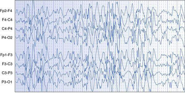
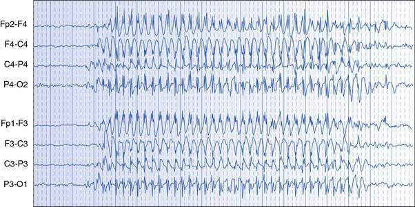
Fig. 17.1.2 The EEG of childhood absence epilepsy during an absence seizure, showing a paroxysm of generalized 3-Hz spike–wave activity.
Epilepsies with absence seizures usually present after 4 years of age and in otherwise normal children. There are two main syndromes described. In childhood absence epilepsy (formerly ‘petit mal’ epilepsy), absences begin before 10 years of age, tonic–clonic seizures are rare, the EEG shows runs of regular 3-Hz spike–wave activity, and prognosis for seizure remission is good. In juvenile absence epilepsy, onset of absences is later, sometimes in the teen years, the EEG may show faster and more irregular spike–wave activity, there may be associated tonic–clonic seizures, and prognosis for seizure remission is poorer.
Treatment
EEG is needed to confirm absence seizures and characterize the epileptic syndrome; brain imaging is unnecessary. Sodium valproate, ethosuximide and lamotrigine are the medications used commonly to treat absence seizures. Treatment is usually for 2 years in typical childhood absence epilepsy, with an expectation of seizure remission, and through puberty into the teen years in juvenile absence epilepsy. Rare refractory cases may respond to treatment with a ketogenic diet.
Nadine, a 6-year-old girl, was noted by her parents to frequently ‘blank out’ while sitting at the dinner table and, most recently, to stop walking and talking while shopping with her mother. These episodes were occurring several times a day and seemed to last only a few seconds. Her schoolteacher had not noticed any problems. Hyperventilation in the clinic room provoked a typical episode lasting 12 seconds, during which Nadine was seen to stop hyperventilating, be unresponsive, fidget with her shirt and have slight bobbing of her eyes. Childhood absence epilepsy was confirmed with an EEG, which showed 3-Hz generalized spike–wave activity during spontaneous and hyperventilation-induced absence seizures. Sodium valproate was introduced slowly over 3 weeks, with no absences noted after the second week of treatment and none precipitated with hyperventilation when reviewed. Slight irritability and moodiness were reported by Nadine’s parents as potential side-effects of treatment.

Stay updated, free articles. Join our Telegram channel

Full access? Get Clinical Tree