Scrotal Pain
Eva Ilse Rubio, MD
DIFFERENTIAL DIAGNOSIS
Common
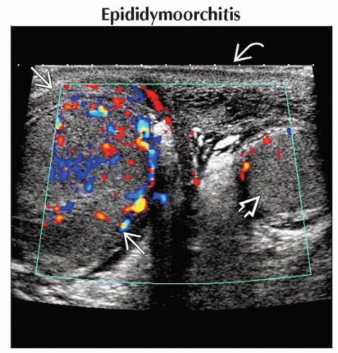
Epididymoorchitis
Testicular Torsion
Inguinal Hernia
Torsion of Testicular/Epididymal Appendage
Varicocele
Hydrocele, Primary or Secondary
Less Common
Primary Neoplasm: Germ Cell Tumors, Stromal Cell Tumors
Metastases: Lymphoma, Leukemia, Neuroblastoma
ESSENTIAL INFORMATION
Key Differential Diagnosis Issues
Specific duration/timing of pain
Known prior medical history
Helpful Clues for Common Diagnoses
Epididymoorchitis
Epididymis/testis often enlarged, hypoechoic/heterogeneous, hypervascular
Reactive hydrocele often present
If recurrent, consider congenital anomaly
Testicular Torsion
Appearance depends on duration
Acute: Enlarged, hypoechoic, absent flow
Subacute/chronic: Heterogeneous, hydrocele, or peritesticular hyperemia
Inguinal Hernia
Loops of bowel in scrotum usually peristalse, though not always
Hernia contents can be mesentery only
Torsion of Testicular/Epididymal Appendage
May involve appendix testis, appendix epididymis, paradidymis
US appearance
Small, echogenic or hypoechoic knot
Hyperemia/hydrocele may mimic epididymitis
Varicocele
Tubular anechoic structures with blood flow on color Doppler with Valsalva
Usually left sided, occasionally bilateral
If unilateral on right, search for underlying venous obstruction
Hydrocele, Primary or Secondary
May be simple fluid or complex with septations, debris
Helpful Clues for Less Common Diagnoses
Primary Neoplasms: Germ Cell Tumors, Stromal Cell Tumors
US characteristics nonspecific
Focal or diffuse abnormality
Hydrocele or hyperemia may be present
Hematoma after minor trauma: Follow to resolution to exclude underlying lesion
Metastases: Leukemia, Lymphoma, Neuroblastoma
US characteristics nonspecific
Focal or diffuse abnormality
May be unilateral or bilateral
Image Gallery
 Transverse color Doppler ultrasound shows an enlarged, painful right testis
 with diffuse scrotal wall thickening with diffuse scrotal wall thickening  and exuberant blood flow. The left testis is normal and exuberant blood flow. The left testis is normal  . .Stay updated, free articles. Join our Telegram channel
Full access? Get Clinical Tree
 Get Clinical Tree app for offline access

|