Chapter 154 Scleroderma and Raynaud Phenomenon
Juvenile scleroderma encompasses a range of conditions unified by the presence of fibrosis of the skin. Juvenile scleroderma is divided into 2 major categories, localized scleroderma (LS, also known as morphea), which is largely limited to the skin, and systemic sclerosis (SSc), with organ involvement. Although localized disease is the predominant type seen in pediatric populations, systemic sclerosis is associated with severe morbidity and mortality.
Etiology and Pathogenesis
The etiology of scleroderma is unknown, but the mechanism of disease appears to be a combination of a vasculopathy, autoimmunity, immune activation, and fibrosis. Triggers, including trauma, infection, and, possibly, subclinical graft versus host reaction from persistent maternal cells (microchimerism), injure vascular endothelial cells, resulting in increased expression of adhesion molecules. These molecules entrap platelets and inflammatory cells, resulting in vascular changes with manifestations such as Raynaud phenomenon and pulmonary hypertension. Inflammatory cells infiltrate the area of initial vascular damage, causing further vascular damage and resulting in thickened artery walls and reduction in capillary numbers. Macrophages and other inflammatory cells then migrate into affected tissues and secrete cytokines that induce fibroblasts to reproduce and synthesize excessive amounts of collagen, resulting in fibrosis and subsequent lipoatrophy, dermal fibrosis, and loss of sweat glands and hair follicles. In late stages, the entire dermis may be replaced by compact collagen fibers.
Autoimmunity is believed to be a key process in the pathogenesis of both localized and systemic scleroderma, given the high percentage of affected children with autoantibodies. Children with localized disease often have a positive ANA test result (42%), and 47% of this subgroup have antihistone antibodies. Other autoantibodies seen include rheumatoid factor (RF) (16%) and antiphospholipid antibodies (12%). The relationship between specific autoantibodies and the various forms of scleroderma is not well understood, and all antibody test results may be negative in a child, especially one who has LS.
Classification
Localized scleroderma is distinct from systemic scleroderma and rarely progresses to systemic disease. Within the category of LS there are several subtypes that are differentiated by both the distribution of the lesions and the depth of involvement (Table 154-1). Up to 15% of children have a combination of 2 or more subtypes.
Table 154-1 CLASSIFICATION OF PEDIATRIC SCLERODERMA (MORPHEA)
LOCALIZED SCLERODERMA
Plaque Morphea
Generalized Morphea
Bullous Morphea
Bullous lesions that can occur with any of the subtypes of morphea
Linear Scleroderma
Linear lesions can extend through the dermis, subcutaneous tissue, and muscle to underlying bone; more likely unilateral
Deep Morphea
SYSTEMIC SCLEROSIS
Diffuse
Limited
Epidemiology
Juvenile scleroderma is rare, with an estimated prevalence of 1/100,000. Localized scleroderma is far more common than SSc in children, by a 10 : 1 ratio, with plaque morphea and linear scleroderma being the most common subtypes. Linear scleroderma is predominantly a pediatric condition, with 65% of patients diagnosed before age 18. After age 8 yr the female : male ratio for both LS and SSc is approximately 3 : 1, whereas in patients younger than 8 yr there is no sex predilection.
Clinical Manifestations
Localized Scleroderma
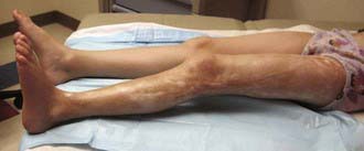
The onset of scleroderma is generally insidious, and manifestations vary according to disease subtype. The initial skin manifestations of localized disease usually include erythema or a bluish hue seen around an area of waxy induration; subtle erythema may be the only presenting sign (Fig. 154-1). Early edema and erythema are followed by indurated, hypopigmented or hyperpigmented, atrophic lesions (Fig. 154-2). Linear scleroderma varies in size from a few centimeters to the entire length of the extremity, with varying depth. Patients sometimes present with arthralgias, synovitis, or flexion contractures (Fig. 154-3). Children also experience limb length discrepancies as a result of growth impairment due to involvement of muscle and bone. Children with en coup de sabre (Fig. 154-4) may have symptoms unique to central nervous system (CNS) involvement, such as seizures, hemifacial atrophy, ipsilateral uveitis, and learning/behavioral changes.

Figure 154-1 Boy with generalized morphea. Note the active circular lesion (arrowheads) with a surrounding rim of erythema. The largest lesion has areas of postinflammatory hyperpigmentation and depression with an area of erythema on the right. The small lesion (arrow) demonstrates depression due to lipoatrophy.

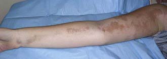
Figure 154-2 Inactive linear scleroderma demonstrating hyperpigmented lesion with areas of normal skin (skip lesions).

Full access? Get Clinical Tree

