Right Ventricle Enlargement
Alexander J. Towbin, MD
DIFFERENTIAL DIAGNOSIS
Common
Pulmonary Hypertension (PH)
Atrial Septal Defect (ASD)
Less Common
Dilated Cardiomyopathy
Pulmonary Stenosis
Pulmonary Thromboembolism (PTE)
Rare but Important
Tricuspid Regurgitation
ESSENTIAL INFORMATION
Key Differential Diagnosis Issues
Causes: ↑ pulmonary vascular resistance, ↑ right heart blood volume
Helpful Clues for Common Diagnoses
Pulmonary Hypertension (PH)
In neonatal period, persistent PH of newborn is most common cause of PH
Association: Neonatal lung disorders → increased pulmonary vascular resistance
Causes of PH in children: Congenital heart defect (CHD) and pulmonary disease
Occurs in CHD when pulmonary blood flow or vascular resistance is ↑
Main pulmonary artery is larger than aorta
Atrial Septal Defect (ASD)
2nd most common isolated CHD
3 types: Secundum (92.5%), primum, and sinus venosus
MR: Differentiated from normal wall thinning by thickening at edge of defect
Helpful Clues for Less Common Diagnoses
Dilated Cardiomyopathy
Typical dilated ventricles & ↓ contractility
Often presents with heart failure
Multiple etiologies: Viral myocarditis, CHD, Takayasu arteritis, metabolic disorders, and nutritional deficiencies
Most often thought to be idiopathic
CXR: Cardiomegaly and pulmonary congestion
Pulmonary Stenosis
Types: Valvular, subvalvular, supravalvular, or in branch pulmonary arteries
Valvular most common (7-9% of all CHD)
CXR: Dilated main pulmonary artery
Pulmonary Thromboembolism (PTE)
Uncommon in children
Risk factors: Central venous lines, malignancy, CHD, lupus, vascular malformations, or renal disease
30-60% have deep vein thrombosis
Chronic PTE → right heart failure and PH
Helpful Clues for Rare Diagnoses
Tricuspid Regurgitation
Can be seen as part of Ebstein anomaly or as isolated anomaly
Isolated tricuspid regurgitation can be caused by papillary muscle rupture
Rare cause in children
Papillary rupture can occur in utero
Image Gallery
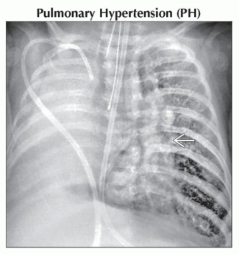
 AP radiograph of the chest shows complete consolidation of the right hemithorax with a rightward shift of the heart and mediastinum due to pulmonary hypoplasia. The left hilum is enlarged
 . .Stay updated, free articles. Join our Telegram channel
Full access? Get Clinical Tree
 Get Clinical Tree app for offline access

|