Rickets and Hypervitaminosis D

Chapter 48 Rickets and Hypervitaminosis D


Rickets


Bone consists of a protein matrix called osteoid and a mineral phase, principally composed of calcium and phosphate, mostly in the form of hydroxyapatite. Osteomalacia is present when there is inadequate mineralization of bone osteoid and occurs in children and adults. Rickets is a disease of growing bone that is due to unmineralized matrix at the growth plates and occurs in children only before fusion of the epiphyses. Because growth plate cartilage and osteoid continue to expand but mineralization is inadequate, the growth plate thickens. There is also an increase in the circumference of the growth plate and the metaphysis, increasing bone width at the location of the growth plates and causing some of the classic clinical manifestations, such as widening of the wrists and ankles. There is a general softening of the bones that causes them to bend easily when subject to forces such as weight bearing or muscle pull. This softening leads to a variety of bone deformities.


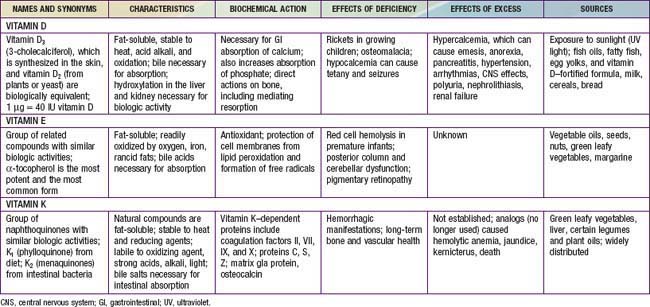
Rickets is principally due to vitamin D deficiency (Table 48-1) and was rampant in northern Europe and the USA during the early years of the 20th century. Although this problem was largely corrected through public health measures that provided children with adequate vitamin D, rickets remains a persistent problem in developed countries, with many cases still secondary to preventable nutritional vitamin D deficiency It remains a significant problem in developing countries as well, with some community-based and general hospital-based surveys among children in Africa finding the prevalence of rickets exceeds 10%. UNICEF has estimated that up to 25% of children in China have some evidence of rickets.


Clinical Manifestations


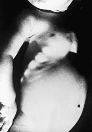
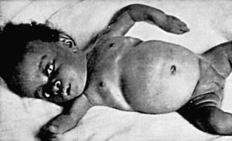
Most manifestations of rickets are due to skeletal changes (Table 48-3). Craniotabes is a softening of the cranial bones and can be detected by applying pressure at the occiput or over the parietal bones. The sensation is similar to the feel of pressing into a Ping-Pong ball and then releasing. Craniotabes may also be secondary to osteogenesis imperfecta, hydrocephalus, and syphilis. It is a normal finding in many newborns, especially near the suture lines, but it typically disappears within a few months of birth. Widening of the costochondral junctions results in a rachitic rosary, which feels like the beads of a rosary as the examiner’s fingers move along the costochondral junctions from rib to rib (Fig. 48-1). Growth plate widening is also responsible for the enlargement at the wrists and ankles. The horizontal depression along the lower anterior chest known as Harrison groove occurs from pulling of the softened ribs by the diaphragm during inspiration (Fig. 48-2). Softening of the ribs also impairs air movement and predisposes patients to atelectasis and pneumonia.


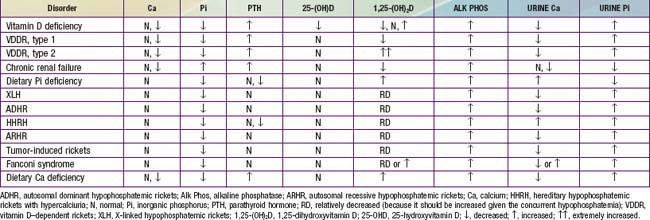
There is some variation in the clinical presentation of rickets based on the etiology. Changes in the lower extremities tend to be the dominant feature in X-linked hypophosphatemic rickets. Symptoms secondary to hypocalcemia occur only in those forms of rickets associated with decreased serum calcium (Table 48-4).


The chief complaint in a child with rickets is quite variable. Many children present because of skeletal deformities, whereas others have difficulty walking owing to a combination of deformity and weakness. Other common presenting complaints include failure to thrive and symptomatic hypocalcemia (Chapter 565).
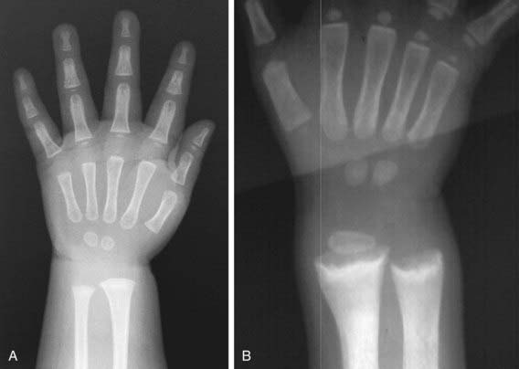
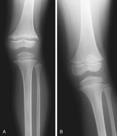


Radiology


Rachitic changes are most easily visualized on posteroanterior radiographs of the wrist, although characteristic rachitic changes can be seen at other growth plates (Figs. 48-3 and 48-4). Decreased calcification leads to thickening of the growth plate. The edge of the metaphysis loses its sharp border, which is described as fraying. The edge of the metaphysis changes from a convex or flat surface to a more concave surface. This change to a concave surface is termed cupping and is most easily seen at the distal ends of the radius, ulna, and fibula. There is widening of the distal end of the metaphysis, corresponding to the clinical observation of thickened wrists and ankles as well as the rachitic rosary. Other radiologic features include coarse trabeculation of the diaphysis and generalized rarefaction.


Clinical Evaluation


Because the majority of children with rickets have a nutritional deficiency, the initial evaluation should focus on a dietary history, emphasizing intake of vitamin D and calcium. Most children in industrialized nations receive vitamin D from formula, fortified milk, or vitamin supplements. Along with the amount, the exact composition of the formula or milk is pertinent, because rickets has occurred in children given products that are called milk (soy milk) but are deficient in vitamin D and/or minerals.


Cutaneous synthesis mediated by sunlight exposure is an important source of vitamin D. It is important to ask about time spent outside, sunscreen use, and clothing, especially if there may be a cultural reason for increased covering of the skin. Because winter sunlight is ineffective at stimulating cutaneous synthesis of vitamin D, the season is an additional consideration. Children with increased skin pigmentation are at increased risk for vitamin D deficiency because of decreased cutaneous synthesis.


The presence of maternal risk factors for nutritional vitamin D deficiency, including diet and sun exposure, is an important consideration when a neonate or young infant has rachitic findings, especially if the infant is breast-fed. Determining a child’s intake of dairy products, the main dietary source of calcium, provides a general sense of calcium intake. High dietary fiber can interfere with calcium absorption.


The child’s medication use is relevant, because certain medications such as the anticonvulsants phenobarbital and phenytoin increase degradation of vitamin D, and aluminum-containing antacids interfere with the absorption of phosphate.


Malabsorption of vitamin D is suggested by a history of liver or intestinal disease. Undiagnosed liver or intestinal disease should be suspected if the child has gastrointestinal (GI) symptoms, although occasionally rickets is the presenting complaint. Fat malabsorption is often associated with diarrhea or oily stools, and there may be signs or symptoms suggesting deficiencies of other fat-soluble vitamins (A, E, and K; Chapters 45, 49, and 50).


A history of renal disease (proteinuria, hematuria, urinary tract infections) is an additional significant consideration, given the importance of chronic renal failure as a cause of rickets. Polyuria can occur in children with chronic renal failure or Fanconi syndrome.


Children with rickets might have a history of dental caries, poor growth, delayed walking, waddling gait, pneumonia, and hypocalcemic symptoms.


The family history is critical, given the large number of genetic causes of rickets, although most of these causes are rare. Along with bone disease, it is important to inquire about leg deformities, difficulties with walking, or unexplained short stature, because some parents may be unaware of their diagnosis. Undiagnosed disease in the mother is not unusual in X-linked hypophosphatemia. A history of a unexplained sibling death during infancy may be present in the child with cystinosis, the most common cause of Fanconi syndrome in children.


The physical examination focuses on detecting manifestations of rickets (see Table 48-3). It is important to observe the child’s gait, auscultate the lungs to detect atelectasis or pneumonia, and plot the patient’s growth. Alopecia suggests vitamin D-dependent rickets type 2.


The initial laboratory tests in a child with rickets should include serum calcium, phosphorus, alkaline phosphatase, parathyroid hormone (PTH), 25-hydroxyvitamin D, 1,25-dihydroxyvitamin D3, creatinine, and electrolytes (see Table 48-4 for interpretation). Urinalysis is useful for detecting the glycosuria and aminoaciduria (positive dipstick for protein) seen with Fanconi syndrome. Evaluation of urinary excretion of calcium (24 hr collection for calcium or calcium:creatinine ratio) is helpful if hereditary hypophosphatemic rickets with hypercalciuria or Fanconi syndrome is suspected. Direct measurement of other fat-soluble vitamins (A, E, and K) or indirect assessment of deficiency (prothrombin time for vitamin K deficiency) is appropriate if malabsorption is a consideration.


Vitamin D Disorders


Vitamin D Physiology


Vitamin D can be synthesized in skin epithelial cells and therefore technically is not a vitamin. Cutaneous synthesis is normally the most important source of vitamin D and depends on the conversion of 7-dehydrochlesterol to vitamin D3 (3-cholecalciferol) by ultraviolet B radiation from the sun. The efficiency of this process is decreased by melanin; hence, more sun exposure is necessary for vitamin D synthesis in people with increased skin pigmentation. Measures to decrease sun exposure, such as covering the skin with clothing or applying sunscreen, also decrease vitamin D synthesis. Children who spend less time outside have reduced vitamin D synthesis. The winter sun away from the equator is ineffective at mediating vitamin D synthesis.


There are few natural dietary sources of vitamin D. Fish liver oils have a high vitamin D content. Other good dietary sources include fatty fish and egg yolks. Most children in industrialized countries receive vitamin D via fortified foods, especially formula and milk (both of which contain 400 IU/L) and some breakfast cereals and breads. Supplemental vitamin D may be vitamin D2 (which comes from plants or yeast) or vitamin D. Breast milk has a low vitamin D content, approximately 12-60 IU/L.


Vitamin D is transported bound to vitamin D–binding protein to the liver, where 25-hydroxlase converts vitamin D into 25-hydroxyvitamin D (25-D), the most abundant circulating form of vitamin D. Because there is little regulation of this liver hydroxylation step, measurement of 25-D is the standard method for determining a patient’s vitamin D status. The final step in activation occurs in the kidney, where 1α-hydroxylase adds a second hydroxyl group, resulting in 1,25-dihydroxyvitamin D (1,25-D). The 1α-hydroxylase is upregulated by PTH and hypophosphatemia; hyperphosphatemia and 1,25-D inhibit this enzyme. Most 1,25-D circulates bound to vitamin D–binding protein.


1,25-D acts by binding to an intracellular receptor, and the complex affects gene expression by interacting with vitamin D–response elements. In the intestine, this binding results in a marked increase in calcium absorption, which is highly dependent on 1,25-D. There is also an increase in phosphorus absorption, but this effect is less significant because most dietary phosphorus absorption is vitamin D independent. 1,25-D also has direct effects on bone, including mediating resorption. 1,25-D directly suppresses PTH secretion by the parathyroid gland, thus completing a negative feedback loop. PTH secretion is also suppressed by the increase in serum calcium mediated by 1,25-D. 1,25-D inhibits its own synthesis in the kidney and increases the synthesis of inactive metabolites.


Nutritional Vitamin D Deficiency


Vitamin D deficiency remains the most common cause of rickets globally and is prevalent, even in industrialized countries. Because vitamin D can be obtained from dietary sources or from cutaneous synthesis, most patients in industrialized countries have a combination of risk factors that lead to vitamin D deficiency.


Etiology


Vitamin D deficiency most commonly occurs in infancy due to a combination of poor intake and inadequate cutaneous synthesis. Transplacental transport of vitamin D, mostly 25-D, typically provides enough vitamin D for the 1st 2 mo of life unless there is severe maternal vitamin D deficiency. Infants who receive formula receive adequate vitamin D, even without cutaneous synthesis. Because of the low vitamin D content of breast milk, breast-fed infants rely on cutaneous synthesis or vitamin supplements. Cutaneous synthesis can be limited due to the ineffectiveness of the winter sun in stimulating vitamin D synthesis; avoidance of sunlight due to concerns about cancer, neighborhood safety, or cultural practices; and decreased cutaneous synthesis because of increased skin pigmentation.


The effect of skin pigmentation explains why most cases of nutritional rickets in the USA and northern Europe occur in breast-fed children of African descent or other dark-pigmented populations. The additional impact of the winter sun is supported by the fact that such infants more commonly present in the late winter or spring. In some groups, complete covering of infants or the practice of not taking infants outside has a significant role, explaining the occurrence of rickets in infants living in areas of abundant sunshine, such as the Middle East. Because the mothers of some infants can have the same risk factors, decreased maternal vitamin D can also contribute, both by leading to reduced vitamin D content in breast milk and by lessening transplacental delivery of vitamin D. Rickets caused by vitamin D deficiency can also be secondary to unconventional dietary practices, such as vegan diets that use unfortified soy milk or rice milk.


Clinical Manifestations


The clinical features are typical of rickets (see Table 48-3), with a significant minority presenting with symptoms of hypocalcemia; prolonged laryngospasm is occasionally fatal. These children have an increased risk of pneumonia and muscle weakness leading to a delay in motor development.


Jun 18, 2016 | Posted by in PEDIATRICS | Comments Off on Rickets and Hypervitaminosis D

Full access? Get Clinical Tree

Get Clinical Tree app for offline access