Retroperitoneal Mass
Alexander J. Towbin, MD
DIFFERENTIAL DIAGNOSIS
Common
Lymphoma
Neuroblastoma
Less Common
Abscess
Lymphatic Malformation
Metastases
Retroperitoneal Hematoma
Duodenal Hematoma
Rare but Important
Neurofibromatosis Type 1
Extraadrenal Pheochromocytoma
Lipoblastoma
Retroperitoneal Fibrosis
ESSENTIAL INFORMATION
Key Differential Diagnosis Issues
Most retroperitoneal masses are malignant
Lymphoma and neuroblastoma most common
US is useful screening modality
Helpful Clues for Common Diagnoses
Lymphoma
3rd most common pediatric malignancy after leukemia and CNS tumors
10-15% of all childhood cancers
Incidence increases with age
Non-Hodgkin lymphoma is more common than Hodgkin lymphoma in children younger than 10 years old
More common in males
Burkitt and diffuse large B-cell lymphoma most common types to affect retroperitoneum
Neuroblastoma
Most common solid extracranial malignancy
6-10% of all childhood cancers
15% of pediatric cancer deaths
4th most common pediatric malignancy (leukemia, CNS tumors, and lymphoma)
2nd most common abdominal neoplasm (Wilms tumor)
> 90% of patients diagnosed before age 5
Median age at diagnosis is 22 months
Peak incidence in 1st year of life (30%)
Can arise anywhere along sympathetic chain
˜ 70% originate in retroperitoneum
35% in adrenal medulla
30-35% in extraadrenal paraspinal ganglia
Mediastinum is 3rd most common location (20%)
Patients < 1 year old have better prognosis
Abdominal mass is most common presentation
Can present with bruising around eyes
Paraneoplastic syndromes in ˜ 2%
50% have metastases at diagnosis
Most common to liver, bone, and bone marrow
Hepatic metastases can be diffuse or nodular
Calcifications are present in ˜ 85%
I-123 MIBG uptake in 90-95% of patients
MR is useful to see intraspinal involvement
Prognosis varies depending on stage
Staged by International Neuroblastoma Staging System
Helpful Clues for Less Common Diagnoses
Abscess
Can be due to ruptured appendix, surgery, Crohn disease, and osteomyelitis
Most infections are polymicrobial
Can cross boundaries
Consider tuberculosis if vertebral osteomyelitis extends to retroperitoneal soft tissues
Lymphatic Malformation
a.k.a. mesenteric cyst
Congenital benign tumor
Can be found at all anatomic locations
Most common in head and neck
˜ 20% in abdomen
˜ 5% in retroperitoneum
Can cross anatomic compartments
Large, thin-walled, multiseptated cystic mass
Rare calcifications in wall
Metastases
Testicular metastases are most common
Spreads to retroperitoneal lymph nodes
Retroperitoneal Hematoma
Can be seen after trauma or in hypocoagulable state
Appearance depends on time between hemorrhage and imaging
Duodenal Hematoma
Unusual finding in setting of trauma
Accounts for < 5% of intraabdominal injuries
Due to forces that compress duodenum between spine and fixed object
Seatbelt, handlebars, and abuse are most common causes
Iatrogenic trauma from instrumentation can also occur
Associated with pancreatic injuries
Helpful Clues for Rare Diagnoses
Neurofibromatosis Type 1
Autosomal dominant disorder
Classical clinical findings include café-au-lait spots, axillary freckling, and dermal and plexiform neurofibromas
Plexiform neurofibromas can occur in abdomen
Most common in abdominal wall and retroperitoneum
Retroperitoneal neurofibromas can cause mass effect on spinal cord, bowel obstruction, and ureteric obstruction
Plexiform neurofibromas have targetoid appearance on MR
Loss of targetoid appearance should raise concern for degeneration into malignant peripheral nerve sheath tumor
Extraadrenal Pheochromocytoma
a.k.a. paraganglioma
˜ 30% of all pediatric pheochromocytomas are extraadrenal
85% of extraadrenal pheochromocytomas are retroperitoneal
Organ of Zuckerkandl is most common site of origin
Most common in 2nd-3rd decade
Associated with von Hippel Lindau, MEN type 2, and neurofibromatosis type 1
20-50% are malignant
Most commonly metastasize to bone, liver, and lungs
Often presents with hypertension
Cause of hypertension in ˜ 1% of children
Appear hyperintense on T2WIs
I-123 MIBG both sensitive and specific for detection
No proven risk of hypertensive crisis with iodinated contrast
Lipoblastoma
Most common in children under age 3
Most commonly occurs in trunk and extremities
Retroperitoneal Fibrosis
Rare disorder in children
In children, 50% are related to systemic process or autoimmune disorder
Insidious onset with nonspecific signs and symptoms
Appears as misty mesentery on CT early in disease process
Image Gallery
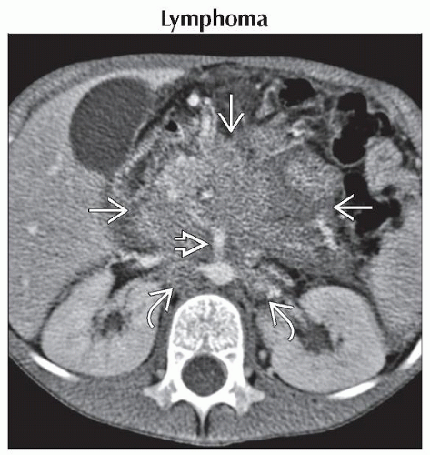 Axial CECT shows a conglomerate nodal mass  surrounding the superior mesenteric artery surrounding the superior mesenteric artery  . Nodes are also present in the periaortic and aortocaval region . Nodes are also present in the periaortic and aortocaval region  . . |
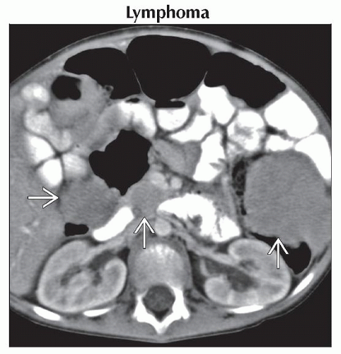 Axial CECT shows multiple enlarged lymph nodes
 . Lymphoma is the 3rd most common malignancy in children, though more common in males than in females. . Lymphoma is the 3rd most common malignancy in children, though more common in males than in females.Stay updated, free articles. Join our Telegram channel
Full access? Get Clinical Tree
 Get Clinical Tree app for offline access

|