Respiratory Tract Disorders

Chapter 95 Respiratory Tract Disorders


Respiratory disorders are the most frequent cause of admission for neonatal intensive care in both term and preterm infants. Signs and symptoms of respiratory distress include cyanosis, grunting, nasal flaring, retractions, tachypnea, decreased breath sounds with or without rales and/or rhonchi, and pallor. A wide variety of pathologic lesions may be responsible for respiratory disturbances, including pulmonary, airway, cardiovascular, central nervous, and other disorders (Fig. 95-1).
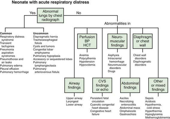


It is occasionally difficult to distinguish respiratory from nonrespiratory etiologies on the basis of clinical signs alone. Any sign of respiratory distress is an indication for a physical examination and diagnostic evaluation, including a blood gas or pulse oximetry determination and radiograph of the chest. Timely and appropriate therapy is essential to prevent ongoing injury and improve outcome. As a result of important advances in treatment respiratory disease as well as in understanding of its pathophysiology, neonatal and infant deaths from early respiratory disease have declined markedly. The challenge is not only to continue to improve survival but also to reduce short- and long-term complications related to early lung disease.


95.1 Transition to Pulmonary Respiration


Waldemar A. Carlo


Successful establishment of adequate lung function at birth depends on airway patency, functional lung development, and maturity of respiratory control. Fetal lung fluid must be removed and replaced with gas. This process begins before birth as active sodium transport across the pulmonary epithelium drives liquid from the lung lumen into the interstitium with subsequent absorption into the vasculature. Increased levels of circulating catecholamines, vasopressin, prolactin, and glucocorticoids enhance lung fluid adsorption and trigger the change in lung epithelia from a chloride-secretory to a sodium-reabsorptive mode. Functional residual capacity (FRC) must be established and maintained in order to develop a ventilation-perfusion relationship that will provide optimal exchange of oxygen and carbon dioxide between alveoli and blood (Chapter 415).


The First Breath


During vaginal delivery, intermittent compression of the thorax facilitates removal of lung fluid. Surfactant lining the alveoli enhances the aeration of gas-free lungs by reducing surface tension, thereby lowering the pressure required to open alveoli. Although spontaneously breathing infants do not need to generate an opening pressure to create airflow, infants requiring positive pressure ventilation at birth need an opening pressure of 13-32 cm H2O and are more likely to establish FRC if they generate a spontaneous, negative pressure breath. Expiratory esophageal pressures associated with the 1st few spontaneous breaths in term newborns range from 45 to 90 cm H2O. This high pressure, due to expiration against a partially closed glottis, may aid in the establishment of FRC but would be difficult to mimic safely with use of artificial ventilation. The higher pressures needed to initiate respiration are required to overcome the opposing forces of surface tension (particularly in small airways) and the viscosity of liquid remaining in the airways, as well as to introduce about 50 mL/kg of air into the lungs, 20-30 mL/kg of which remains after the 1st breath to establish FRC. Air entry into the lungs displaces fluid, decreases hydrostatic pressure in the pulmonary vasculature, and increases pulmonary blood flow. The greater blood flow, in turn, increases the blood volume of the lung and the effective vascular surface area available for fluid uptake. The remaining fluid is removed via the pulmonary lymphatics, upper airway, mediastinum, and pleural space. Fluid removal may be impaired after cesarean section or as a result of surfactant deficiency, endothelial cell damage, hypoalbuminemia, high pulmonary venous pressure, or neonatal sedation.


Initiation of the 1st breath is due to a decline in PaO2 and pH and a rise in PaCO2 as a result of interruption of the placental circulation, a redistribution of cardiac output, a decrease in body temperature, and various tactile and sensory inputs. The relative contributions of these stimuli to the onset of respiration are uncertain.


When compared with term infants, the low birthweight (LBW) infant who has a very compliant chest wall may be at a disadvantage in establishing FRC. The FRC is lowest in the most immature infants because of the decrease in alveolar number. Abnormalities in ventilation-perfusion ratio are greater and persist for longer periods in LBW infants and may lead to hypoxemia and hypercarbia as a result of atelectasis, intrapulmonary shunting, hypoventilation, and gas trapping. The smallest immature infants have the most profound disturbances, which may resemble respiratory distress syndrome (RDS). However, even in healthy term infants, oxygenation is impaired soon after birth, and oxygen saturation improves to exceed 90% at around 5 minutes.


95.2 Apnea


Apnea is a common problem in preterm infants that may be due to prematurity or an associated illness. In term infants, apnea is always worrisome and demands immediate diagnostic evaluation. Periodic breathing must be distinguished from prolonged apneic pauses, because the latter may be associated with serious illnesses. Apnea is a feature of many primary diseases that affect neonates (Table 95-1). These disorders produce apnea by direct depression of the central nervous system’s control of respiration (hypoglycemia, meningitis, drugs, hemorrhage, seizures), disturbances in oxygen delivery (shock, sepsis, anemia), or ventilation defects (obstruction of the airway, pneumonia, muscle weakness).


Table 95-1 POTENTIAL CAUSES OF NEONATAL APNEA AND BRADYCARDIA


Central nervous system Intraventricular hemorrhage, drugs, seizures, hypoxic injury, herniation, neuromuscular disorders, Leigh syndrome, brainstem infarction or anomalies (e.g., olivopontocerebellar atrophy), after general anesthesia
Respiratory Pneumonia, obstructive airway lesions, upper airway collapse, atelectasis, extreme prematurity, laryngeal reflex, phrenic nerve paralysis, pneumothorax, hypoxia
Infectious Sepsis, meningitis (bacterial, fungal, viral), respiratory syncytial virus, pertussis
Gastrointestinal Oral feeding, bowel movement, necrotizing enterocolitis, intestinal perforation
Metabolic ↓ Glucose, ↓ calcium, ↓/↑ sodium, ↑ ammonia, ↑ organic acids, ↑ ambient temperature, hypothermia
Cardiovascular Hypotension, hypertension, heart failure, anemia, hypovolemia, vagal tone
Other Immaturity of respiratory center, sleep state

Idiopathic apnea of prematurity occurs in the absence of identifiable predisposing diseases. Apnea is a disorder of respiratory control and may be obstructive, central, or mixed. Obstructive apnea (pharyngeal instability, neck flexion) is characterized by absence of airflow but persistent chest wall motion. Pharyngeal collapse may follow the negative airway pressures generated during inspiration, or it may result from incoordination of the tongue and other upper airway muscles involved in maintaining airway patency. In central apnea, which is caused by decreased central nervous system (CNS) stimuli to respiratory muscles, airflow and chest wall motion are absent. Gestational age is the most important determinant of respiratory control, with the frequency of apnea being inversely related to gestational age. The immaturity of the brainstem respiratory centers is manifested by an attenuated response to carbon dioxide and a paradoxical response to hypoxia that results in apnea rather than the hyperventilation observed after the 1st months of life. The most common pattern of idiopathic apnea in preterm neonates is mixed apnea (50-75% of cases), with obstructive apnea preceding (usually) or following central apnea. Short episodes of apnea are usually central, whereas prolonged ones are often mixed. Apnea depends on the sleep state; its frequency increases during active (rapid eye movement) sleep.


Treatment


Infants at risk for apnea should be started on cardiorespiratory monitoring. Gentle tactile stimulation is often adequate therapy for mild and intermittent episodes. The onset of apnea in a previously well premature neonate after the 2nd wk of life or in a term infant at any time is a critical event that warrants immediate investigation. Recurrent apnea of prematurity may be treated with theophylline or caffeine. Methylxanthines increase central respiratory drive by lowering the threshold of response to hypercapnia as well as enhancing contractility of the diaphragm and preventing diaphragmatic fatigue. Theophylline and caffeine are as effective, but caffeine has fewer side effects (tachycardia, feeding intolerance). Loading doses of 5-7 mg/kg of theophylline (orally) or aminophylline (intravenously) should be followed by doses of 1-2 mg/kg given every 6-12 hr by the oral or intravenous route. Loading doses of 20 mg/kg of caffeine citrate are followed 24 hr later by maintenance doses of 5 mg/kg/24 hr qd, either orally or intravenously. These doses should be monitored through observation of vital signs and clinical response. Serum drug determinations (therapeutic levels: theophylline, 6-10 µg/mL; caffeine, 8-20 µg/mL) are optional because important side effects of these agents are rare. Higher doses of methylxanthines are more effective, do not result in frequent side effects, and tend to reduce major neurodevelopmental disabilities. Withholding respiratory stimulants in infants with RDS may result in ventilator dependency, increased bronchopulmonary dysplasia (BPD), and death. Doxapram, known to be a potent respiratory stimulant, acts predominantly on peripheral chemoreceptors and is effective in neonates with apnea of prematurity that is unresponsive to methylxanthines. Transfusion of packed red blood cells to reduce the incidence of idiopathic apnea is reserved for severely anemic infants. Gastroesophageal reflux is common in neonates, but data do not support a causal relationship between gastroesophageal reflux and apneic events or the use of antireflux medications to reduce the frequency of apnea in preterm infants.


Nasal continuous positive airway pressure (continuous positive airway pressure [CPAP], 2-5 cm H2O) and high-flow humidification using nasal cannula (1-2.5 L/min) are therapies for mixed or obstructive apnea, but CPAP is preferred because of its proven efficacy and safety. The efficacy of CPAP is related to its ability to splint the upper airway and prevent airway obstruction.


Bibliography


Arad-Cohen N, Cohen A, Tirosh E. The relationship between gastroesophageal reflux and apnea in infants. J Pediatr. 2000;137:321-326.


Committee on Fetus and Newborn, American Academy of Pediatrics. Apnea, sudden infant death syndrome, and home monitoring. Pediatrics. 2003;111:914-917.


Kimball AL, Carlton DP. Gastroesophageal reflux medications in the treatment of apnea in premature infants. J Pediatr. 2001;138:355-360.


Litmanovitz I, Carlo WA. Expectant management of pneumothorax in ventilated neonates. Pediatrics. 2008;122:e975-e979.


Martin RJ, Abu-Shaweesh JM, Baird TM. Apnoea of prematurity. Paediatr Respir Rev. 2004;5:S377-S382.


Ramanathan R, Corwin MJ, Hunt CE, et al. Cardiorespiratory events recorded on home monitors: comparison of healthy infants with those at increased risk for SIDS. JAMA. 2001;285:2199-2207.


Schmidt B, Roberts RS, Davis P, et al. Long term effects of caffeine for apnea of prematurity. N Engl J Med. 357, 2007. 1983–1902


Schmidt B, Roberts RS, Davis P, et al. Caffeine therapy for apnea of prematurity. N Engl J Med. 2006;354:2112-2120.


Sreenan C, Lemke RP, Hudson-Mason A, et al. High-flow nasal cannulae in the management of apnea of prematurity: a comparison with conventional nasal continuous positive airway pressure. Pediatrics. 2001;107:1081-1083.


Steer P, Flenady V, Shearman A, et al. High dose caffeine citrate for extubation of preterm infants: a randomized controlled trial. Arch Dis Child Fetal Neonatal Ed. 2004;89:F499-F503.


Tauman R, Sivan Y. Duration of home monitoring for infants discharged with apnea of prematurity. Biol Neonate. 2000;78:168-173.


95.3 Respiratory Distress Syndrome (Hyaline Membrane Disease)


Etiology and Pathophysiology


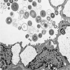
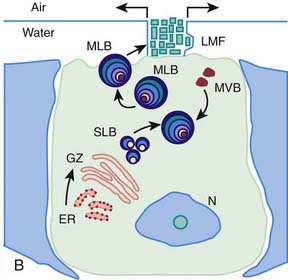
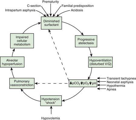
Surfactant deficiency (decreased production and secretion) is the primary cause of RDS. The failure to attain an adequate FRC and the tendency of affected lungs to become atelectatic correlate with high surface tension and the absence of pulmonary surfactant. The major constituents of surfactant are dipalmitoyl phosphatidylcholine (lecithin), phosphatidylglycerol, apoproteins (surfactant proteins SP-A, SP-B, SP-C, and SP-D), and cholesterol (Fig. 95-2). With advancing gestational age, increasing amounts of phospholipids are synthesized and stored in type II alveolar cells (Fig. 95-3). These surface-active agents are released into the alveoli, where they reduce surface tension and help maintain alveolar stability by preventing the collapse of small air spaces at end-expiration. Because of immaturity, the amounts produced or released may be insufficient to meet postnatal demands. Surfactant is present in high concentrations in fetal lung homogenates by 20 wk of gestation, but it does not reach the surface of the lungs until later. It appears in amniotic fluid between 28 and 32 wk. Mature levels of pulmonary surfactant are present usually after 35 wk. Though rare, genetic disorders may contribute to respiratory distress. Abnormalities in surfactant protein B and C genes as well as a gene responsible for transporting surfactant across membranes (ABC transporter 3 [ABCA3]) are associated with severe and often lethal familial respiratory disease. Other familial causes of neonatal respiratory distress (not RDS) include alveolar capillary dysplasia, acinar dysplasia, pulmonary lymphangiectasia, and mucopolysaccharidosis.


Figure 95-3 A, Fetal rat lung (low magnification), day 20 (term, day 22) showing developing type II cells, stored glycogen (pale areas), secreted lamellar bodies, and tubular myelin.


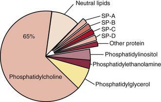
(Courtesy of Mary Williams, MD, University of California, San Francisco.)

image

Synthesis of surfactant depends in part on normal pH, temperature, and perfusion. Asphyxia, hypoxemia, and pulmonary ischemia, particularly in association with hypovolemia, hypotension, and cold stress, may suppress surfactant synthesis. The epithelial lining of the lungs may also be injured by high oxygen concentrations and the effects of respirator management, thereby resulting in a further reduction in surfactant.


Alveolar atelectasis, hyaline membrane formation, and interstitial edema make the lungs less compliant in RDS, so greater pressure is required to expand the alveoli and small airways. In affected infants, the lower part of the chest wall is pulled in as the diaphragm descends, and intrathoracic pressure becomes negative, thus limiting the amount of intrathoracic pressure that can be produced; the result is the development of atelectasis. The chest wall of the preterm infant, which is highly compliant, offers less resistance than that of the mature infant to the natural tendency of the lungs to collapse. Thus, at end-expiration, the volume of the thorax and lungs tends to approach residual volume, and atelectasis may develop.


Deficient synthesis or release of surfactant, together with small respiratory units and a compliant chest wall, produces atelectasis and results in perfused but not ventilated alveoli, causing hypoxia. Decreased lung compliance, small tidal volumes, increased physiologic dead space, and insufficient alveolar ventilation eventually result in hypercapnia. The combination of hypercapnia, hypoxia, and acidosis produces pulmonary arterial vasoconstriction with increased right-to-left shunting through the foramen ovale and ductus arteriosus and within the lung itself. Pulmonary blood flow is reduced and ischemic injury both to the cells producing surfactant and to the vascular bed results in an effusion of proteinaceous material into the alveolar spaces (Fig. 95-4).


Clinical Manifestations


Signs of RDS usually appear within minutes of birth, although they may not be recognized for several hours in larger premature infants until rapid, shallow respirations have increased to 60 breaths/min or greater. A late onset of tachypnea should suggest other conditions. Some patients require resuscitation at birth because of intrapartum asphyxia or initial severe respiratory distress (especially with a birthweight < 1,000 g). Characteristically, tachypnea, prominent (often audible) grunting, intercostal and subcostal retractions, nasal flaring, and cyanosis are noted. Breath sounds may be normal or diminished with a harsh tubular quality, and on deep inspiration, fine rales may be heard. The natural course of untreated RDS is characterized by progressive worsening of cyanosis and dyspnea. If the condition is inadequately treated, blood pressure may fall; cyanosis and pallor increase, and grunting decreases or disappears, as the condition worsens. Apnea and irregular respirations are ominous signs requiring immediate intervention. Patients may also have a mixed respiratory-metabolic acidosis, edema, ileus, and oliguria. Respiratory failure may occur in infants with rapid progression of the disease. In most cases, the symptoms and signs reach a peak within 3 days, after which improvement is gradual. Improvement is often heralded by spontaneous diuresis and improved blood gas values at lower inspired oxygen levels and/or lower ventilator support. Death can be due to severe impairment of gas exchange, alveolar air leaks (interstitial emphysema, pneumothorax), pulmonary hemorrhage, or IVH. Death may be delayed by weeks or months if BPD develops in infants with severe RDS.


Diagnosis


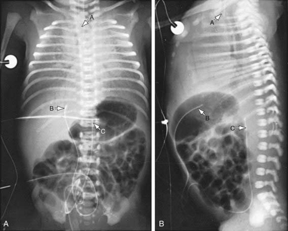
The clinical course, chest radiographic findings, and blood gas and acid-base values help establish the clinical diagnosis. On radiographs, the lungs may have a characteristic but not pathognomonic appearance that includes a fine reticular granularity of the parenchyma and air bronchograms, which are often more prominent early in the left lower lobe because of superimposition of the cardiac shadow (Fig. 95-5). The initial radiographic appearance is occasionally normal, with the typical pattern developing at 6-12 hr. Considerable variation in film findings may be seen, depending on the phase of respiration (inspiratory vs expiratory radiograph) and the use of CPAP or positive end-expiratory pressure (PEEP); this variation often results in poor correlation between radiographic findings and the clinical course. Laboratory findings are characterized initially by hypoxemia and later by progressive hypoxemia, hypercapnia, and variable metabolic acidosis.


In the differential diagnosis, early-onset sepsis may be indistinguishable from RDS. In pneumonia manifested at birth, the chest roentgenogram may be identical to that for RDS. Maternal group B streptococcal colonization, identification of organisms on gram staining of gastric or tracheal aspirates or a buffy coat smear, and/or the presence of marked neutropenia may suggest the diagnosis of early-onset sepsis. Cyanotic heart disease (total anomalous pulmonary venous return) can also mimic RDS both clinically and radiographically. Echocardiography with color-flow imaging should be performed in infants who show no response to surfactant replacement, to rule out cyanotic congenital heart disease as well as ascertain patency of the ductus arteriosus and assess pulmonary vascular resistance (PVR). Persistent pulmonary hypertension, aspiration (meconium, amniotic fluid) syndromes, spontaneous pneumothorax, pleural effusions, and congenital anomalies such as cystic adenomatoid malformation, pulmonary lymphangiectasia, diaphragmatic hernia, and lobar emphysema must be considered in patients with an atypical clinical course but can generally be differentiated from RDS through radiographic evaluation. Transient tachypnea may be distinguished by its short and mild clinical course and is characterized by low or no need for oxygen supplementation. Congenital alveolar proteinosis (congenital surfactant protein B deficiency) is a rare familial disease that manifests as severe and lethal RDS in predominantly term and near-term infants (Chapter 399). In atypical cases of RDS, a lung profile (lecithin:sphingomyelin ratio and phosphatidylglycerol determination) performed on a tracheal aspirate can be helpful in establishing a diagnosis of surfactant deficiency.


Prevention


Avoidance of unnecessary or poorly timed cesarean section, appropriate management of high-risk pregnancy and labor, and prediction of pulmonary immaturity with possible in utero acceleration of maturation (Chapter 90) are important preventive strategies. In timing of cesarean section or induction of labor, estimation of fetal head circumference by ultrasonography and determination of the lecithin concentration in amniotic fluid by the lecithin:sphingomyelin ratio (particularly useful with phosphatidylglycerol measurement in diabetic pregnancies) decrease the likelihood of delivering a premature infant. Antenatal and intrapartum fetal monitoring may similarly decrease the risk of fetal asphyxia; asphyxia is associated with an increased incidence and severity of RDS.


Administration of antenatal corticosteroids to women between 24 and 34 wk of gestation significantly reduces the incidence and mortality of RDS as well as overall neonatal mortality. Antenatal steroids also reduce (1) the need for and duration of ventilatory support and admission to a neonatal intensive care unit (NICU) and (2) the incidence of severe IVH, necrotizing enterocolitis, early-onset sepsis, and developmental delay. Postnatal growth is not adversely affected. Antenatal steroids do not increase the risk of maternal death, chorioamnionitis, or puerperal sepsis. Corticosteroid administration is recommended for all women in preterm labor (24-34 wk of gestation) who are likely to deliver a fetus within 1 wk. Repeated weekly doses of betamethasone until 32 wk may reduce neonatal morbidities and the duration of mechanical ventilation. Antenatal glucocorticoids act synergistically with postnatal exogenous surfactant therapy so they should be given even though surfactant therapy is so effective. Betamethasone and dexamethasone have been used antenatally. Dexamethasone may result in a lower incidence of IVH than betamethasone, but further research is needed to determine whether one of these steroids is superior for antenatal treatment.


Administration of a 1st dose of surfactant into the trachea of symptomatic premature infants immediately after birth (prophylactic) or during the 1st few hours of life (early rescue) reduces air leak and mortality from RDS but does not alter the incidence of BPD.


Treatment


The basic defect requiring treatment in RDS is inadequate pulmonary exchange of oxygen and carbon dioxide; metabolic acidosis and circulatory insufficiency are secondary manifestations. Early supportive care of premature infants, especially in the treatment of acidosis, hypoxia, hypotension (Chapter 92), and hypothermia, may lessen the severity of RDS. Therapy requires careful and frequent monitoring of heart and respiratory rates, oxygen saturation, PaO2, PaCO2, pH, serum bicarbonate, electrolytes, glucose, and hematocrit, blood pressure, and temperature. Arterial catheterization is frequently necessary. Because most cases of RDS are self-limited, the goal of treatment is to minimize abnormal physiologic variations and superimposed iatrogenic problems. Treatment of infants with RDS is best carried out in the NICU.


The general principles for supportive care of any premature infant should be adhered to, including developmental care and scheduled “touch times.” To avoid hypothermia and minimize oxygen consumption, the infant should be placed in an incubator or radiant warmer, and core temperature maintained between 36.5 and 37°C (Chapters 91 and 92). Use of an incubator is preferable in very LBW (VLBW) infants owing to the high insensible water losses associated with radiant heat. Calories and fluids should initially be provided intravenously. For the 1st 24 hr, 10% glucose and water should be infused through a peripheral vein at a rate of 65-75 mL/kg/24 hr. Electrolytes should be added on day 2 in the most mature infants and on days 3 to 7 in the more immature ones. Fluid volume is increased gradually over the 1st week. Excessive fluids (> 140 mL/kg/day) contribute to the development of patent ductus arteriosus (PDA) and BPD.


Warm humidified oxygen should be provided at a concentration initially sufficient to keep arterial oxygen pressure between 40 and 70 mm Hg (85-95% saturation) in order to maintain normal tissue oxygenation while minimizing the risk of oxygen toxicity. If oxygen saturation cannot be kept > 85% at inspired oxygen concentrations of 40-70% or greater, applying CPAP at a pressure of 5-10 cm H2O via nasal prongs is indicated and usually produces a sharp improvement in oxygenation. CPAP prevents collapse of surfactant-deficient alveoli and improves both FRC and ventilation-perfusion matching. Early use of CPAP for stabilization of at-risk VLBW infants beginning as early as in the delivery room reduces ventilatory needs. Another approach is to intubate the VLBW infant, administer intratracheal surfactant, and then extubate the infant and begin CPAP. The amount of CPAP required usually decreases after about 72 hr of age, and most infants can be weaned from CPAP shortly thereafter. If an infant with RDS undergoing CPAP cannot keep oxygen saturation > 85% while breathing 40-70% oxygen, assisted ventilation and surfactant are indicated.


Infants with respiratory failure or persistent apnea require assisted mechanical ventilation. Reasonable measures of respiratory failure are: (1) arterial blood pH <7.20, (2) arterial blood PCO2 of 60 mmHg or higher, and (3) oxygen saturation <85% at oxygen concentrations of 40-70% and CPAP of 5-10 cm H2O. Infants with persistent apnea also need mechanical ventilation. Intermittent positive pressure ventilation delivered by time-cycled, pressure-limited, continuous flow ventilators is a common method of conventional ventilation for newborns. Other methods of conventional ventilation are synchronized intermittent mandatory ventilation (the set rate and pressure synchronized with the patient’s own breaths), pressure support (the patient triggers each breath and a set pressure is delivered), and volume ventilation (a mode in which a specific tidal volume is set and the delivered pressure varies), and combinations thereof. Assisted ventilation for infants with RDS should always include PEEP (Chapter 65.1). High ventilatory rates (60/min) result in fewer air leaks. With use of high ventilatory rates, sufficient expiratory time should be allowed to avoid the administration of inadvertent PEEP.


The goal of mechanical ventilation is to improve oxygenation and elimination of carbon dioxide without causing pulmonary injury or oxygen toxicity. Acceptable ranges of blood gas values, after the risks of hypoxia and acidosis are balanced against those of mechanical ventilation, vary among institutions: PaO2 40-70 mmHg, PaCO2 45-65 mmHg, and pH 7.20-7.35. During mechanical ventilation, oxygenation is improved by increasing either the fraction of inspired oxygen (FIO2) or the mean airway pressure. The latter can be increased by raising the peak inspiratory pressure, PEEP gas flow, or inspiratory-expiratory ratio. Pressure changes are usually most effective. However, excessive PEEP may impede venous return, thereby reducing cardiac output and decreasing oxygen delivery despite improvement in PaO2. PEEP levels of 4-6 cm H2O are usually safe and effective. Carbon dioxide elimination is achieved by increasing the peak inspiratory pressure (tidal volume) or the rate of the ventilator.


A strategy to minimize ventilator-associated lung injury is the use of CPAP instead of endotracheal intubation. The decreased need for ventilator support with the use of CPAP may allow lung inflation to be maintained but may prevent volutrauma due to overdistention and/or atelectasis. However, controlled trials do not report benefits of early CPAP. Interestingly, nasal intermittent mandatory ventilation (vs nasal CPAP) reduces extubation failure in small trials; this method could be an alternative by which to avoid intubation.


The strategy most evaluated with conventional mechanical ventilation is the use of high rates and presumably small tidal volumes as PaCO2 levels were kept in comparable ranges. Meta-analyses of the randomized controlled trials comparing high (>60 per min) and low (usually 30-40 per min) rates (and presumed low vs. high tidal volumes, respectively) revealed that the high ventilatory rate strategy led to fewer air leaks and a trend for increased survival.


If mechanical ventilation is needed, a ventilatory approach using small tidal volumes and permissive hypercapnia can be employed. Permissive hypercapnia is a strategy for the management of patients receiving ventilatory support in which priority is given to the prevention or limitation of lung injury from the ventilator by tolerating relatively high levels of PaCO2 rather than maintenance of normal blood gas values. A multicenter trial of infants ≤1,000 g reported that permissive hypercapnia (target PaCO2 >50 mm Hg) during the 1st 10 days led to a trend for lower rates of BPD or death at 36 wk. Furthermore, the strategy of permissive hypercapnia reduced severity of BPD, as evidenced by a decreased need for ventilator support at 36 wk, from 16% to 1%. A large multi-center randomized controlled trial of permissive hypercapnia with emphasis on the use of CPAP revealed that this is an effective and maybe preferred approach to the standard strategy of intubation and surfactant administration to preterm infants with RDS. Volume-targeted ventilation allows the clinician to set a tidal volume that may prevent volutrama. There are limited data on volume-targeted ventilation, but this mode of ventilation may decrease the rates of pneumothorax and BPD.


Hyperoxia may also contribute to lung injury in preterm infants. Thus, permissive hypoxemia is another strategy that may reduce BPD. Trials to target different levels of oxygen saturation performed for treatment of retinopathy of prematurity or BPD revealed that the groups with lower saturation targets (89-94% or 91-94%, respectively) had less need for oxygen supplementation and lower rates of BPD or BPD exacerbation. Even lower oxygen saturation targets (85-89%) reduced retinopathy of prematurity significantly and tended to reduce BPD but increased mortality rates in a large multi-center randomized controlled trial. Thus, optimal target saturations are still not determined but some restriction of oxygen appears beneficial.


Many ventilated neonates receive sedation or pain relief with benzodiazepines or opiates (morphine, fentanyl), respectively. Midazolam is approved for use in neonates and has demonstrated sedative effects. Adverse hemodynamic effects and myoclonus have been associated with its use in neonates. If it is used, a continuous infusion or administration of individual doses over at least 10 min is recommended to reduce these risks. Data are insufficient to assess the efficacy and safety of lorazepam. Diazepam is not recommended owing to its long half-life, its long-acting metabolites, and concern about the benzyl alcohol content of diazepam injection. Continuous infusion of morphine in VLBW neonates requiring mechanical ventilation does not reduce mortality rates, severe IVH, or periventricular leukomalacia (PVL). The need for additional doses of morphine is associated with poor outcome.


High-frequency ventilation (HFV) achieves desired alveolar ventilation by using smaller tidal volumes and higher rates (300-1,200 breaths/min or 5-20 Hz). HFV may improve the elimination of carbon dioxide and improve oxygenation in patients who show no response to conventional ventilators and who have severe RDS, interstitial emphysema, recurrent pneumothoraces, or meconium aspiration pneumonia. High-frequency oscillatory ventilation (HFOV) and high-frequency jet ventilation (HFJV) are the most frequently used methods of HFV. HFOV reduces BPD but increases air leaks and may raise the risk for IVH and PVL. HFOV strategies that promote lung recruitment, combined with surfactant therapy, may improve gas exchange. HFJV facilitates resolution of air leaks. Elective use of either method, in comparison with conventional ventilation, generally does not offer advantages if used as the initial ventilation strategy to treat infants with RDS.


Surfactant deficiency is the primary pathophysiology of RDS. Immediate effects of surfactant replacement therapy include improved alveolar-arterial oxygen gradients, reduced ventilatory support, increased pulmonary compliance, and improved chest radiograph appearance . Treatment is initiated as soon as possible in the hours after birth. Repeated dosing is given via the endotracheal tube every 6-12 hr for a total of 2 to 4 doses, depending on the preparation. Exogenous surfactant should be given by a physician who is qualified in neonatal resuscitation and respiratory management and who is able to care for the infant beyond the 1st hr of stabilization. Additional on-site staff support required includes nurses and respiratory therapists experienced in the ventilatory management of premature infants. Appropriate monitoring equipment (radiology, blood gas laboratory, pulse oximetry) must also be available. Complications of surfactant therapy include transient hypoxia, hypercapnia, bradycardia and hypotension, blockage of the endotracheal tube, and pulmonary hemorrhage (Chapter 95.13).


A number of surfactant preparations are available, including synthetic surfactants and natural surfactants derived from animal sources. Exosurf is a synthetic surfactant. Natural surfactants include Survanta (bovine), Infasurf (calf), and Curosurf (porcine). Surfactant replacement therapy is one of the major advances in the care of preterm infants. Prophylactic and rescue administrations of synthetic and natural surfactants have reduced adverse outcomes, including mortality. Specifically, neonatal mortality is lower with prophylactic (vs rescue) administration of both synthetic and natural surfactants. Prophylactic administration of both types of surfactants decreases the risk for pneumothorax and pulmonary interstitial emphysema. The lack of reduction in BPD rates following surfactant replacement is probably, in part, due to the survival of infants with severe RDS who would have died without surfactant administration.


In more mature preterm infants, a policy of prophylactic administration of surfactant may result in many infants’ receiving surfactant unnecessarily, and it is possible that early rather than prophylactic surfactant administration may be sufficiently effective. However, the time of rescue surfactant administration in the prophylactic trials varied widely. Early (< 2 hr) rather than delayed rescue surfactant administration resulted in decreased risk for neonatal mortality. Early rescue administration of surfactant reduced rates of both pneumothorax (from 14% to 12%) and pulmonary interstitial emphysema (from 15% to 10%). An alternative to early surfactant administration in many infants is to treat infants with surfactant before ventilation is needed. Temporary endotracheal intubation for surfactant administration in infants requiring only CPAP reduces the subsequent need for mechanical ventilation and may reduce mortality and/or BPD.


Head-to-head trials of natural and synthetic surfactants report superiority of the natural surfactants. Use of natural (vs synthetic) surfactants resulted in lower rates of pneumothorax (12% vs 7%) and mortality (18% vs. 16%) than use of synthetic surfactants. Natural surfactants are superior because of their surfactant-associated protein content, their more rapid onset, and their lower risk of pneumothorax and improved survival. Surfaxin, formerly known as KL4 surfactant, is a novel synthetic lung surfactant containing phospholipids and an engineered peptide, sinapultide, designed to mimic the actions of human SP-B. Use of Surfaxin for the prevention and treatment of RDS demonstrates equivalency to use of the natural surfactants Survanta and Curosurf. Initial protocols for surfactant administration used single-dose therapies. However, compared with a strategy of a single dose of surfactant, administration of multiple doses of surfactant when indicated according to the protocol resulted in lower pneumothorax risk (18% vs 9%) and a trend for lower mortality. A review of all the current evidence supports the use of prophylactic or early use of natural surfactants as early as when infants require CPAP. More than one dose of surfactant should be administered if indicated to optimize the benefits of this therapy.


Premature infants requiring ventilator support after 1 wk of age experience transient episodes of surfactant dysfunction associated with deficiencies of SP-B and SP-C, which are temporally associated with episodes of infection and respiratory deterioration.


Inhaled nitric oxide (iNO) decreases the need for extracorporeal membrane oxygenation (ECMO) in term and near-term infants with hypoxic respiratory failure or persistent pulmonary hypertension of the neonate. The response to iNO is equivalent to that to HFOV in term or near-term infants with hypoxic respiratory failure. A positive response to combined therapy suggests that alveolar recruitment by HFOV may allow iNO gas to reach the pulmonary resistance vessels. A reduction in the rate of death or BPD in infants <1,000 g treated with iNO was observed in one study but not in others.


Strategies for weaning infants from ventilators vary widely and are influenced by lung mechanics as well as the availability of ventilatory modes (pressure support). Once extubated, many infants are transitioned to nasal CPAP to avoid postextubation atelectasis and reduce re-intubation. Synchronized nasal intermittent ventilation decreases the need for re-intubation in premature infants. High flow (1-2 L/min) or warmed, humidified high-flow (2-8 L/min) nasal cannula oxygen is commonly used to support term and near-term infants following extubation and to wean premature infants from nasal CPAP. Preloading with methylxanthines may enhance the success of extubation.


Pharmacologic Therapies


Several pharmacologic options are available to the clinician for the treatment of RDS and prevention of its complications. Selected treatments are reviewed here.


Vitamin A supplementation given largely to infants < 1,000 g resulted in a decrease in death and/or BPD at 36 wk (from 66% to 60%) and trends for less nosocomial sepsis and retinopathy of prematurity.


Systemic corticosteroids have been used to treat infants with RDS, to selectively treat infants who continue to require respiratory support, and to treat those in whom BPD develops. Mortality and/or BPD at 36 wk decrease (from 72% to 45%) with moderately early (7-14 days) administration of corticosteroids. Early (<96 hr) and delayed (> 2-3 wk) administration of systemic steroids has also been assessed with meta-analyses, and the results are qualitatively similar. However, there are short-term adverse effects, including hyperglycemia, hypertension, gastrointestinal bleeding, gastrointestinal perforation, hypertrophic obstructive cardiomyopathy, poor weight gain, poor growth of the head, and a trend toward a higher incidence of PVL. Furthermore, data showing an increased incidence of neurodevelopmental delay and cerebral palsy in infants randomly assigned to receive systemic corticosteroids raise serious concerns about adverse long-term outcomes of this therapy. Thus, routine use of systemic corticosteroids for the prevention or treatment of BPD is not recommended by the Consensus Group of the American Academy of Pediatrics and the Canadian Pediatric Society. Administration of inhaled steroids to ventilated preterm infants during the 1st 2 wk after birth reduced the need for systemic steroids (from 45% to 35%) and tended to decrease rates of death and/or BPD at 36 wk without an increase in adverse effects.


Inhaled NO has been evaluated in preterm infants following the observation of its effectiveness in term and near-term infants with hypoxemic respiratory failure. Despite optimistic results from a large randomized controlled trial, trials in preterm infants report heterogeneous effects on BPD, mortality, and other important outcomes. The most current data do not support the routine administration of iNO in preterm infants with hypoxemic respiratory failure.


Prevention of extubation failure has been attempted with use of various pharmacologic approaches. Methylaxanthines appear to have a large effect on reducing extubation (from 51% to 25%). Similarly, use of systemic steroids before extubation reduces the need for re-intubation (from 10% to 1%). In contrast, administration of racemic epinephrine after extubation does not improve pulmonary function or the rate of extubation failure.


Metabolic acidosis in RDS may be a result of perinatal asphyxia and hypotension and is often encountered when an infant has required resuscitation (Chapter 94). Sodium bicarbonate, 1-2 mEq/kg, may be administered over 15-20 min through a peripheral or umbilical vein, followed by an acid-base determination within 30 min, or it may be administered over several hours. Often, sodium bicarbonate is administered on an emergency basis through an umbilical venous catheter. Alkali therapy may result in skin slough from infiltration, increased serum osmolarity, hypernatremia, hypocalcemia, hypokalemia, and liver injury when concentrated solutions are administered rapidly through an umbilical vein catheter wedged in the liver.


Monitoring of aortic blood pressure through an umbilical or peripheral arterial catheter or by oscillometric technique is useful in managing the shock-like state that may occur during the 1st hr or so in premature infants who have been asphyxiated or have severe RDS (see Fig. 94-2). The position of a radiopaque umbilical catheter should be checked roentgenographically after insertion (see Fig. 95-5). The tip of an umbilical artery catheter should lie just above the bifurcation of the aorta (L3-L5) or above the celiac axis (T6-T10). Preferred sites for peripheral catheters are the radial or posterior tibial arteries. The placement and supervision should be carried out by skilled and experienced personnel. Catheters should be removed as soon as patients no longer have any indication for their continued use—usually when an infant is stable and the FIO2 is < 40%. Hypotension and low flow in the superior vena cava (SVC) have been associated with higher rates of CNS morbidity and mortality and should be treated with cautious administration of volume (crystalloid) and early use of vasopressors. Dopamine is more effective in raising blood pressure than dobutamine. Hypotension may be refractory to pressors, but responsive to glucocorticoids, especially in neonates < 1,000 g. This hypotension may be due to transient adrenal insufficiency in the ill premature infant. It should be treated with intravenous hydrocortisone (Solu-Cortef) at 1-2 mg/kg/dose q6-12 hr (Chapter 92).


Periodic monitoring of PaO2, PaCO2, and pH is an important part of the management; if assisted ventilation is being used, such monitoring is essential. Oxygenation may be assessed continuously from transcutaneous electrodes or pulse oximetry (oxygen saturation). Capillary blood samples are of limited value for determining PO2 but may be useful for evaluating PCO2 and pH.


Because of the difficulty of distinguishing group B streptococcal or other bacterial infections from RDS, empirical antibiotic therapy is indicated until the results of blood cultures are available. Penicillin or ampicillin with an aminoglycoside is suggested, although the choice of antibiotics should be based on the recent pattern of bacterial sensitivity in the hospital where the infant is being treated (Chapter 103).


Jun 18, 2016 | Posted by in PEDIATRICS | Comments Off on Respiratory Tract Disorders

Full access? Get Clinical Tree

Get Clinical Tree app for offline access