CHAPTER 18 Radiographic assessment of adult brachial plexus injuries
Summary box
Introduction
Despite the many advances in microsurgical strategies and techniques during the last several decades, treatment of brachial plexus injuries remains challenging. Of paramount importance to surgical planning is a thorough characterization of the injury affecting various elements of the brachial plexus. For instance, accurate detection of nerve root avulsion is crucial. If the nerve is traumatically transected distal to the dorsal root ganglion (postganglionic), nerve repair using sural nerve grafts remains a viable option for restoration of motor function. In contrast, if the lesion is proximal to the dorsal root ganglion (preganglionic), nerve repair is uncommon, and nerve transfer (neurotization) becomes the more useful strategy.1
Nerve root avulsions complicate more than 70% of traumatic brachial plexus injuries, so what are the most reliable diagnostic techniques used to define the nature of the lesion(s)? Clinical examination cannot routinely distinguish between pre- and postganglionic injuries, although the presence of Horner’s sign suggests proximal injury to lower elements of the brachial plexus. Electrodiagnostic examinations contribute to accurate diagnosis of brachial plexus injuries, but again the information is inferred,2 not directly demonstrated. On the other hand, radiographic assessments are beginning to provide methods for visualizing the anatomic defect. In the setting of traumatic injury to the brachial plexus, radiographic studies can be most useful in the detection of preganglionic injuries3 and will be the focus of this chapter.
The advent of magnetic resonance imaging (MRI) greatly augmented the diagnostic workup of traumatic brachial plexus injuries. For many decades, identification of avulsed nerve roots relied solely on invasive examinations such as myelography and computed tomographic (CT) myelography (CTM). The increasing sophistication in MRI techniques now offers additional diagnostic modalities such as MR myelography (MRM), which has completely replaced invasive myelographic examinations at some institutions. Another alternative method for the radiographic assessment of brachial plexus injuries employs the technique of MR neurography (MRN).4 However, in our opinion, CTM and, more recently, MRM, remain the most reliable tools for radiographic assessment and should be performed as part of the routine diagnostic/preoperative evaluation of patients with brachial plexus injuries.
Myelography
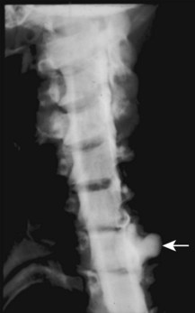
Radiographic detection of nerve root avulsion in brachial plexus injury was essentially a chance discovery. In 1947, Murphey et al.5 performed myelography on a patient with severe pain and a brachial plexus injury in order to exclude the presence of a concomitant herniated cervical disk. The resultant myelogram demonstrated mushroom-shaped leakages of iodinated contrast material bulging from the dural sac extending beyond the vertebral foramina (Box 18.1). In 1949, the same author described similar myelographic examinations on 7 patients, 3 of which submitted to a surgical exploration that revealed undeniable nerve root avulsions. Subsequently, myelography became routinely used for the diagnosis of nerve root avulsion in brachial plexus palsy.
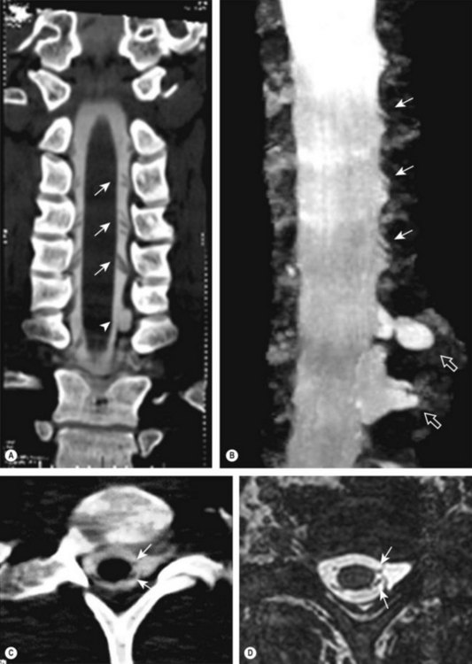
Several myelographic patterns have been described to accurately reflect the corresponding anatomic lesions. The most common is the traumatic pseudomeningocele, which appears as mushroom-shaped leakage of iodinated contrast medium bulging from the dural sac, which may extend beyond the vertebral foramen (Figure 18.1). The vast majority of nerve root avulsions are associated with traumatic pseudomeningoceles, yet rootlet avulsions in the absence of dural abnormalities and pseudomeningoceles have been well-documented (Figure 18.2 A–D).6 Therefore, the presence of a pseudomeningocele strongly suggests an avulsion injury, but is no longer regarded as incontrovertible evidence of nerve root avulsion.
A more reliable indicator of nerve root avulsion is the aspect of nerve root shadows within the dural sleeve. Nagano et al.7 studied 90 adult patients with traumatic brachial plexus injuries and correlated their radiographic appearance with intraoperative findings. They reported a wide spectrum of myelographic abnormalities, ranging from slightly abnormal root sleeve shadows (indicating a reduction in number or size of nerve rootlets or partial avulsion) to complete obliteration of the nerve root sleeve tip with complete absence of the filling defect of the nerve root (complete avulsion). The authors concluded that the complete absence of filling defects of the nerve roots within the root sleeve is associated with a preganglionic injury in 96.5% of cases. Additionally, evaluation of nerve rootlets can be assisted by identification of deformities of the subarachnoid spaces, which are often enlarged or irregular when partial or complete avulsions occur.
The importance of a meticulous myelographic evaluation of the individual nerve rootlets has been recently emphasized in a report8 documenting the usefulness of X-ray myelography in traumatic injuries of the brachial plexus. Complete or partial nerve root avulsions can occur in the absence of pseudomeningoceles in approximately 20% of cases visualized by X-ray myelography; however, when this technique is combined with CTM, the overall accuracy of detection increases to 92%. Note that for evaluation of intradural nerve roots, X-ray myelography can be more sensitive in detecting root avulsions at C8 and T1 levels than CTM; however, it has limited value in the identification of partial avulsions, because ventral and dorsal nerve roots cannot be evaluated separately.9
CT myelography
With the advent of CT, CTM has rapidly become the standard examination for traumatic injuries of the brachial plexus (Box 18.2). Compared to standard myelography, CTM requires less intrathecal volume of the water-soluble iodinated contrast medium, which decreases incidence of adverse effects while maintaining high contrast resolution. Other advantages include high spatial resolution, reduced movement artefacts, and excellent visualization of bone structures. Since the introduction of the multidetector CT, a large number of sub-millimeter sections can be acquired in a very short time, thereby facilitating immediate postprocessing of the study. Disadvantages of CTM include increased radiation exposure (compared to standard myelography), occasional artifacts due to beam hardening, and poor visualization of soft tissue elements such as the distal brachial plexus.


