Pyloric Stenosis and Other Congenital Anomalies of the Stomach

Chapter 321 Pyloric Stenosis and Other Congenital Anomalies of the Stomach


321.1 Hypertrophic Pyloric Stenosis


Hypertrophic pyloric stenosis occurs in 1-3/1,000 infants in the United States. It is more common in whites of northern European ancestry, less common in blacks, and rare in Asians. Males (especially first-borns) are affected approximately 4 to 6 times as often as females. The offspring of a mother and, to a lesser extent, the father who had pyloric stenosis are at higher risk for pyloric stenosis. Pyloric stenosis develops in approximately 20% of the male and 10% of the female descendants of a mother who had pyloric stenosis. The incidence of pyloric stenosis is increased in infants with B and O blood groups. Pyloric stenosis is occasionally associated with other congenital defects, including tracheoesophageal fistula and hypoplasia or agenesis of the inferior labial frenulum.


Clinical Manifestations


Nonbilious vomiting is the initial symptom of pyloric stenosis. The vomiting may or may not be projectile initially but is usually progressive, occurring immediately after a feeding. Emesis might follow each feeding, or it may be intermittent. The vomiting usually starts after 3 wk of age, but symptoms can develop as early as the 1st wk of life and as late as the 5th mo. About 20% have intermittent emesis from birth that then progresses to the classic picture. After vomiting, the infant is hungry and wants to feed again. As vomiting continues, a progressive loss of fluid, hydrogen ion, and chloride leads to hypochloremic metabolic alkalosis. Greater awareness of pyloric stenosis has led to earlier identification of patients with fewer instances of chronic malnutrition and severe dehydration and at times a subclinical self-resolving hypertrophy.


Hyperbilirubinemia is the most common clinical association of pyloric stenosis, also known as icteropyloric syndrome. Unconjugated hyperbilirubinemia is more common than conjugated and usually resolves with surgical correction. It may be associated with a decreased level of glucuronyl transferase as seen in ∼5% of affected infants; mutations in the bilirubin uridine diphosphate glucuronosyl transferase gene (UGT1A1) have also been implicated. If conjugated hyperbilirubinemia is a part of the presentation, other etiologies need to be investigated. Other coexistent clinical diagnoses have been described, including eosinophilic gastroenteritis, hiatal hernia, peptic ulcer, congenital nephrotic syndrome, congenital heart disease, and congenital hypothyroidism.


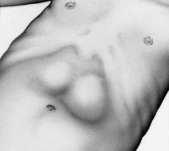
The diagnosis has traditionally been established by palpating the pyloric mass. The mass is firm, movable, ∼2 cm in length, olive shaped, hard, best palpated from the left side, and located above and to the right of the umbilicus in the mid-epigastrium beneath the liver’s edge. The olive is easiest palpated after an episode of vomiting. After feeding, there may be a visible gastric peristaltic wave that progresses across the abdomen (Fig. 321-1).


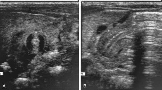
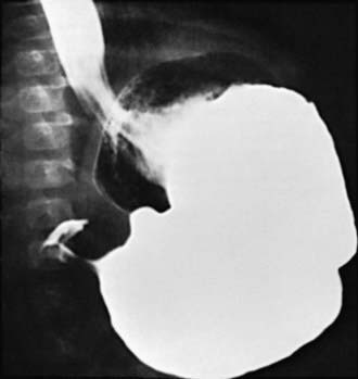
Two imaging studies are commonly used to establish the diagnosis. Ultrasound examination confirms the diagnosis in the majority of cases. Criteria for diagnosis include pyloric thickness 3-4 mm, an overall pyloric length 15-19 mm, and pyloric diameter of 10-14 mm (Fig. 321-2). Ultrasonography has a sensitivity of ∼95%. When contrast studies are performed, they demonstrate an elongated pyloric channel (string sign), a bulge of the pyloric muscle into the antrum (shoulder sign), and parallel streaks of barium seen in the narrowed channel, producing a “double tract sign” (Fig. 321-3).


Jun 18, 2016 | Posted by in PEDIATRICS | Comments Off on Pyloric Stenosis and Other Congenital Anomalies of the Stomach

Full access? Get Clinical Tree

Get Clinical Tree app for offline access