Chapter 118 Primary Defects of Antibody Production
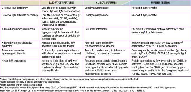
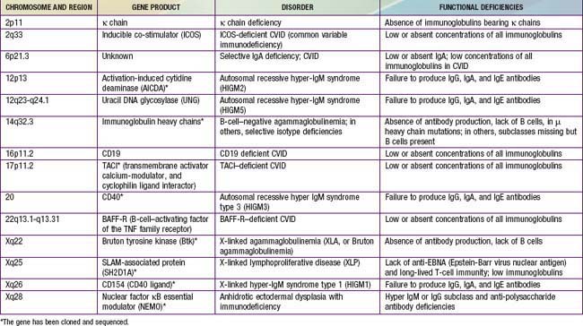
Of all of the primary immunodeficiency diseases, those affecting antibody production are most frequent. Selective absence of serum and secretory IgA is the most common defect, with rates ranging from 1/333 to 1/18,000 persons among different races. By contrast, agammaglobulinemia is estimated to occur with a frequency of only 1/10,000 to 1/50,000 persons. Patients with antibody deficiency are usually recognized because they have recurrent infections with encapsulated bacteria predominantly in the upper and lower respiratory tracts; some individuals with selective IgA deficiency or infants with transient hypogammaglobulinemia may have few or no infections (see Table 116-4). The defective gene products for many primary antibody deficiency disorders have been identified (Table 118-1) and localized (Fig. 118-1). Sometimes the defect is not in the B cell itself but in T cells, which are required for complete B-cell function.

Figure 118-1 Locations of mutant proteins (X) in B cells identified in primary immunodeficiency diseases. β2m, β2 microglobulin; BLNK, B-cell linker adaptor protein; Btk, Bruton tyrosine kinase; HLA, human leukocyte antigen; Ig, immunoglobulin; RFX, RFXAP and CIITA transcription factors; SLAM, signaling lymphocyte activation molecule; TAP1 and TAP2, transporters of processed antigen.
(From Buckley RH: Primary immunodeficiency diseases due to defects in lymphocytes, N Engl J Med 343:1313–1324, 2000.)
X-Linked Agammaglobulinemia (XLA)
Patients with X-linked agammaglobulinemia (XLA), or Bruton agammaglobulinemia, have a profound defect in B-lymphocyte development resulting in severe hypogammaglobulinemia, an absence of circulating B cells, small to absent tonsils, and no palpable lymph nodes.
Genetics and Pathogenesis
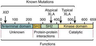
The abnormal gene in XLA maps to q22 on the long arm of the X chromosome and encodes the B-cell protein tyrosine kinase Btk (Bruton tyrosine kinase). Btk is a member of the Tec family of cytoplasmic protein tyrosine kinases and is expressed at high levels in all B-lineage cells, including pre–B cells. It appears to be necessary for pre–B-cell expansion and maturation into surface Ig-expressing B cells, but probably has a role at all stages of B-cell development; it has also been found in cells of the myeloid series. More than 500 different mutations in the human Btk gene are recognized; they encompass most parts of the coding portions of the gene. There is not a clear correlation between the location of the mutation and the clinical phenotype (Fig. 118-2). Carriers are detected by mutation analysis, and prenatal diagnosis of affected male fetuses is possible if the mutation is known in the family.

Figure 118-2 Location of mutations in the functional domains of the Bruton tyrosine kinase (Btk) protein. Deletion and point mutations in Btk identified to date in many boys with classic X-linked agammaglobulinemia (XLA) are in the kinase domain, whereas CBA/N XID mice with a less severe B-cell defect have a point mutation at position 28 in the N-terminal domain. More recently, however, boys with classic XLA are also reported to have mutations at the XID mutation site and in the SH2 domain.
(From Buckley RH: Breakthroughs in the understanding and therapy of primary immunodeficiency, Pediatr Clin North Am 41:665–690, 1994.)
The expression of Btk in cells of myeloid lineage is of interest because boys with XLA often have neutropenia at the height of an acute infection. It is conceivable that Btk is only one of the signaling molecules participating in myeloid maturation and that neutropenia is observed in XLA only when rapid production of such cells is needed. Some pre–B cells are found in the bone marrow; the percentage of peripheral blood B lymphocytes is <1%. The percentage of T cells is increased, ratios of T-cell subsets are normal, and T-cell function is intact. The thymus is normal.
Six autosomal recessive defects have also been shown to result in agammaglobulinemia with an absence of circulating B cells (see Fig. 118-2), including mutations in the genes encoding: (1) the µ heavy chain gene; (2) the Igα and Igβ signaling molecules; (3) B-cell linker adaptor protein (BLNK); (4) the surrogate light chain, λ5/14.1; and (5) leucine-rich repeat-containing 8 (LRRC8).
Clinical Manifestations
Most boys afflicted with XLA remain well during the 1st 6-9 mo of life by virtue of maternally transmitted IgG antibodies. Thereafter, they acquire infections with extracellular pyogenic organisms, such as Streptococcus pneumoniae and Haemophilus influenzae, unless they are given prophylactic antibiotics or immunoglobulin therapy. Infections include sinusitis, otitis media, pneumonia, or, less often, sepsis or meningitis. Infections with Mycoplasma are also particularly problematic. Chronic fungal infections are seen; Pneumocystis jiroveci pneumonia rarely occurs. Viral infections are usually handled normally with the exceptions of hepatitis viruses and enteroviruses. There were several examples of paralysis when live polio vaccine was administered to these patients and chronic, eventually fatal, central nervous system infections with various echoviruses and coxsackieviruses have occurred in a significant number of them. Echovirus-associated myositis resembling dermatomyositis has also been observed. These observations suggest a primary role for antibody, particularly secretory IgA, in host defense against enteroviruses. Growth hormone deficiency has also been reported in association with XLA.
Diagnosis
The diagnosis of XLA should be suspected if lymphoid hypoplasia is found on physical examination (minimal or no tonsillar tissue and no palpable lymph nodes), and serum concentrations of IgG, IgA, IgM, and IgE are far below the 95% confidence limits for appropriate age- and race-matched controls (Chapter 708), usually with total immunoglobulins <100 mg/dL. Levels of natural antibodies to type A and B red blood cell polysaccharide antigens (isohemagglutinins) and antibodies to antigens given during routine immunizations are abnormally low in this disorder, whereas they are normal in transient hypogammaglobulinemia of infancy. Flow cytometry is an important test to demonstrate the absence of circulating B cells, which will distinguish this disorder from common variable immunodeficiency, the hyper-IgM syndrome and transient hypogammaglobulinemia of infancy.
Common Variable Immunodeficiency
Common variable immunodeficiency (CVID) is a syndrome characterized by hypogammaglobulinemia with phenotypically normal B cells. It has also been called “acquired hypogammaglobulinemia” because of a generally later age of onset of infections. CVID patients may appear similar clinically to those with XLA in the types of infections experienced and bacterial etiologic agents involved, except that echovirus meningoencephalitis is rare in patients with CVID (Table 118-2). In contrast to XLA, the sex distribution in CVID is almost equal, the age of onset is later (although it may be present in infancy), and infections may be less severe.
Table 118-2 DIFFERENTIAL DIAGNOSIS FOR PATIENTS WITH SUSPECTED CVID (HYPOGAMMAGLOBULINEMIA WITH RECURRENT INFECTIONS)*

Genetics and Pathogenesis
Most patients have no identified molecular diagnosis. CVID is a category of primary immunodeficiency disorders that likely consists of several different genetic defects with autosomal recessive or dominant inheritance. Genes known to produce the CVID phenotype when mutated include ICOS (inducible co-stimulator) deficiency, SH2DIA (responsible for X-linked lymphoproliferative disease [XLP]), CD19, BAFF-R (B-cell–activating factor of the TNF [tumor necrosis factor] family receptors), and TACI (transmembrane activator, calcium-modulator, and cyclophilin ligand interactor). These mutations in aggregate accounts for less than 10% of all cases of CVID.
Because CVID occurs in 1st-degree relatives of patients with selective IgA deficiency, and some patients with IgA deficiency later become panhypogammaglobulinemic, a large subtype of CVID may have a common genetic basis with IgA deficiency. The high incidence of abnormal immunoglobulin concentrations, autoantibodies, autoimmune disease, and malignancy in both CVID and IgA deficiency and in other members of their families also suggests a shared hereditary influence. This concept is supported by the discovery of a high incidence of C4-A gene deletions and C2 rare gene alleles in the class III major histocompatibility complex (MHC) region in individuals with either IgA deficiency or CVID, suggesting that a common susceptibility gene is on chromosome 6. Only a few human leukocyte antigen (HLA) haplotypes are shared by individuals affected with IgA deficiency and CVID, with at least 1 of 2 particular haplotypes being present in 77% of those affected. In 1 large family with 13 members, 2 had IgA deficiency and 3 had CVID. All of the immunodeficient patients in the family had at least 1 copy of an MHC haplotype that is abnormally frequent in IgA deficiency and CVID: HLA-DQB1 *0201, HLA-DR3, C4B-Sf, C4A-deleted, G11-15, Bf-0.4, C2a, HSP70-7.5, TNFa-5, HLA-B8, and HLA-A1. In a study of 83 multiply affected families with IgA deficiency and CVID, increased allele sharing at chromosome 6p21 in the proximal part of the MHC was observed in a susceptibility locus now designated as IGAD1. More sensitive genetic analysis in 101 multiple-case and 110 single-case families further localized the defect to the HLADQ/DR locus. Environmental factors, particularly drugs such as phenytoin, D-penicillamine, gold, and sulfasalazine are suspected to be triggers for disease expression in individuals with the permissive genetic background.
Most cases of CVID are sporadic or follow an autosomal dominant pattern of inheritance. Patients who lack inducible co-stimulator (ICOS), a surface protein on activated T cells, have an autosomal recessive pattern of inheritance. Nine such patients from 6 families in the Black Forest of Germany have been found to have identical homozygous large genomic deletions of the ICOS gene, suggesting a founder effect. Those who have XLP have an X-linked pattern of inheritance and those with autosomally inherited TACI defects may have heterozygous or homozygous mutations.
Despite normal numbers of circulating immunoglobulin-bearing B lymphocytes and the presence of lymphoid cortical follicles, blood B lymphocytes from CVID patients do not differentiate normally into immunoglobulin-producing cells when stimulated with pokeweed mitogen (PWM) in vitro, even when co-cultured with normal T cells. CVID B cells from some patients can be stimulated both to switch isotype and to synthesize and secrete some immunoglobulin when stimulated with anti-CD40 and IL-4 or IL-10. T cells and T-cell subsets are usually present in normal percentages, although T-cell function is depressed in some patients.
Clinical Manifestations
The serum immunoglobulin and antibody deficiencies in CVID may be as profound as in XLA. Patients with CVID often have autoantibody formation and normal-sized or enlarged tonsils and lymph nodes, and ≈25% of patients have splenomegaly. CVID has also been associated with a sprue-like syndrome with or without nodular follicular lymphoid hyperplasia of the intestine, thymoma, alopecia areata, hemolytic anemia, gastric atrophy, achlorhydria, thrombocytopenia, and pernicious anemia. Lymphoid interstitial pneumonia, pseudolymphoma, B-cell lymphomas, amyloidosis, and noncaseating sarcoid-like granulomas of the lungs, spleen, skin, and liver also occur. There is a 438-fold increase in lymphomas among affected women in the 5th and 6th decades of life. CVID has been reported to resolve transiently or permanently in patients who acquire human immunodeficiency virus (HIV) infection.

Stay updated, free articles. Join our Telegram channel

Full access? Get Clinical Tree

