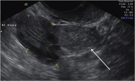
Fig. 3.1
An early intrauterine gestation sac misclassified as a pseudosac
My Management
A.
I agree with the management above
B.
I would proceed to laparoscopy as the hCG is high
C.
I would repeat the transvaginal ultrasound scan
D.
I would carry out uterine curettage
E.
I would repeat the serum hCG
Diagnosis and Assessment
A patient presenting with risk factors for EP (e.g. previous pelvic infection) should heighten suspicion. The greatest risk factor for an EP is tubal damage, which usually occurs after tubal surgery or following a pelvic infection, particularly with Chlamydia trachomatis. Others include smoking and conception following assisted reproduction. However, it must be remembered that most EPs occur in women who do not have any risk factors [1]. Dating a pregnancy by LMP is helpful but not necessarily an accurate predictor of what to expect to see on an ultrasound scan [2]. A significant proportion of women are unsure of their dates or have irregular cycles. Furthermore, we know there are clinically relevant variations in the ovulation–implantation interval [3].
Transvaginal ultrasonography is the best imaging modality for investigating problems in early pregnancy, with > 70 % of EPs visualized at presentation and > 90 % before surgery using this approach [4]. The majority (60 %) of EPs will be identified as a small homogeneous mass (blob sign, Fig. 3.2) rather than the classically described ring or ‘bagel’ sign [5]. The concept of a ‘pseudosac’ is to some extent outdated and relates to a misinterpretation of the ultrasound findings. A round or oval fluid-filled hypoechogenic collection in the uterus in a woman with a positive pregnancy test is more likely to be an early intrauterine gestation sac and should not be cited as evidence for the presence of an EP [6]. Great care should also be taken when performing a scan in women when the uterus is axial or in the presence of fibroids. Both can make it difficult to identify an early intrauterine gestation sac.


Fig. 3.2
A homogeneous mass or ‘blob sign’—the commonest ultrasound feature of an ectopic pregnancy
The case presented had a pregnancy of unknown location (PUL) , not a confirmed EP. The correct management would have been to repeat the transvaginal ultrasound scan at an interval to see if the hypoechogenic area in the endometrial cavity could be definitively identified as an intrauterine pregnancy, or an EP mass visualized before considering methotrexate.
The modern management of PUL is based on assigning risk (low risk: failing PUL and viable intrauterine; high risk: EP) rather than locating the pregnancy. Such risk assessment may be carried using the hCG ratio (hCG at 48 h/hCG at 0 h), although we have recently shown the mathematical prediction model ‘M4’ based on the initial hCG and hCG ratio is effective in this context and outperforms single measurements of progesterone and the hCG ratio alone [7]. This model can be downloaded from: http://homes.esat.kuleuven.be/~biomed/M4PUL/M4triage.htm.
When considering methotrexate administration, it is important to exclude a viable intrauterine pregnancy. Methotrexate can be safely given when an EP has been definitively visualized. However, as most EPs are seen as a homogenous mass [5], false positive test results are possible. Accordingly, waiting 48 hours and reviewing the hCG ratio prior to methotrexate administration can significantly reduce the risk of inadvertent termination. A serum hCG rise of < 35 % is highly unlikely to be associated with a viable intrauterine pregnancy [8]. In this case, the rise in serum hCG of 37.5 % should have alerted the clinical team that the presence of a viable intrauterine pregnancy is potentially possible. A further safeguard prior to methotrexate administration is to ensure a clinician who has specific expertise in early pregnancy imaging has repeated the ultrasound scan.
Historically, it was felt that a viable pregnancy would result in a doubling of serum hCG levels , a failing pregnancy, a halving of serum hCG levels and an EP, a ‘suboptimal rise’ in serum hCG after 48 hours. This does not, however, necessarily aid the clinician in determining the location of the pregnancy as EPs may demonstrate a ‘doubling’ or ‘halving’ of the serum hCG. In contrast, 8 % of viable intrauterine pregnancies have been found to demonstrate a ‘suboptimal rise’ in hCG levels, as seen in this case [8]. In any event, management is based on risk assessment. If the serum hCG is falling, the pregnancy will fall into the low-risk category irrespective of whether it is in the uterine cavity.
Stay updated, free articles. Join our Telegram channel

Full access? Get Clinical Tree


