Fig. 1.1
a Ultrasonographic image of tubal ectopic pregnancy (EP) with both embryonic pole and yolk sac visible within the gestational sac. b Same tubal EP demonstrating cardiac activity using M-Mode (viable EP)
Differential Diagnosis
Ectopic pregnancy
Threatened or incomplete miscarriage
Appendicitis
Urinary tract infection
Ovarian torsion
Pelvic inflammatory disease
Urinary calculus
Gastroenteritis
Ruptured or hemorrhagic ovarian cyst
Our patient had been trying to conceive for the last 6 months and now had a positive urine pregnancy test. Therefore, all the complications of early pregnancy including ectopic and early miscarriage should be considered. A quantitative serum hCG would be helpful to confirm the urine test and also as a tool to estimate the age of pregnancy along with the TVS. More importantly, serial serum hCG levels can be measured as well as TVS examination during the conservative management and follow up of patients diagnosed with EP.
The mild tachycardia and relatively low blood pressure, in addition to abdominal tenderness, peritonitis, blood in vagina, and adnexal tenderness, are indicative of clinical instability and potential rupture of EP and the urgency of diagnosis and management.
Risk Factors for EP
There are several studies published regarding EP and its risk factors. However, only about 50 % of women diagnosed with an EP have identifiable risk factors . Recognition of these risk factors can assist the clinicians not only in the early diagnosis of EP but also in reducing the risk of morbidity and mortality of massive intra-abdominal hemorrhage . Most papers have categorized the risk factors to high, medium, and low risks, although there are variations depending on the epidemiology of the study. Below, we have reviewed the most widely accepted factors for EP. [11, 12].
Previous EP and Tubal Surgery
Previous EP is one of the high risk factors and the incidence increases among people who have had a history of an EP. A woman who has had two prior EPs has a tenfold increased in future EP. This could be due to the tubal dysfunction as the main pathology or secondary to the treatment of EP. The recurrence rate of EP after surgical or nonsurgical management has been reported from 8 to 15 % and 15 % after conservative management [13, 14]. The risk of EP also increases in women who have had a history of any type of pelvic surgery. For example, previous appendectomy increases the risk of EP by twofold [15]. Among the group of women for whom tubal sterilization has failed, pregnancy can result in an EP rate as high as 33 %. Among these patients, the risk of EP is higher in those less than 30 years of age [13].
PID, Infections, and Multiple Sexual Partners
The growing rate of EP is strongly associated with the increasing rate of PID. The incidence of EP increased by more than twofold from 1970 to 1985 from 7 to 16 per 1000 and then declined by 30 % from 1985 to 1997. This was explained by the increase and decline of PID within those periods [16]. It has also been proven that having multiple sexual partners is a strong risk factor for EP with the odds ratio of 2.1 [17]; but the association between PID and number of sexual partners has to be considered [18, 19].
In a European study, 65 % of women with EP had suffered from tubal salpingitis. A history of tubal pathology or tubal surgery has been shown to increase the risk of EP with the odds ratio of 3.8–21.0 and 21.0, respectively [20]. Overall, the history of genital infections, including sexually transmitted disease, PID, and/or any tubal pathology or surgery, is a high risk factor for tubal EP.
Smoking
There are several studies which have confirmed the increased risk of EP in smokers. The risk of EP increases by threefold to fourfold in women who smoke more than one packet of cigarettes per day. The level of risk has been proven to be variable depending on the number of cigarettes smoked. Smoking more than 20 cigarettes a day increases the risk of EP more than smoking 1–5 cigarettes a day with the odds ratio 1.7–3.5 [14, 21].
Infertility
It has been proven that the duration of infertility is associated with increased risk of EP with an adjusted odd ratio of 2.7 for more than 2 years of infertility [21]. The rate of EP is 2–3 % higher in patients undergoing an in vitro fertilization (IVF) [20]. In addition, treatment with gonadotropin and other drugs such as clomiphene in IVF pregnancy increases the incidence of EP. This can also be due to dysfunction of the fallopian tubes [23–26]. The rate of heterotopic pregnancy in the assisted reproductive population could be up to 1 in 100 to 1 in 45 [27, 28].
Other Causes
There are other proven risk factors for EP such as diethylstilbestrol (DES) exposure, intrauterine contraceptive devices, surgical termination of pregnancy, and age. In utero exposure to DES increases the relative risk of EP by 3.84. Intrauterine contraceptive devices (IUCD) such as copper IUCD and Mirena intra-uterine system (IUS) decrease the risk of an EP, but if pregnancy does occur with the device in situ, the risk of EP is higher. Of the 0.5 per 100 Mirena IUS users who become pregnant in 5 years (cumulatively), half are EPs. Regular vaginal douching three to four times per month can increase the risk of PID as a high risk factor for EP by three to four times. Women aged 35–44 years have three times risk of EP compared to younger women. Surgical terminations of pregnancy, spontaneous miscarriages, and older age have all also been shown to increase the risk of EP [11, 21, 29–35].
Types of EP
More than 90–98 % of EPs are tubal pregnancies. EP can also be found in the cervix, ovaries, interstitial tube, cesarean scar, the horn of a bicornuate pregnancy (cornual), or abdomen. About 2–3 % of EPs are implanted in the interstitial portion of the tube (interstitial EP). If the ectopic is implanted in one horn of bicornuate uterus, it is called cornual. Of all EPs, 1 % is cervical, 1–3 % is ovarian, and 1–2 % is abdominal pregnancies. Cesarean scar EP is another rare type of EP, the incidence of which is increasing. [36–40] (Fig. 1.2).
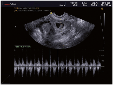
Fig. 1.2
Laparoscopic image of right tubal ectopic pregnancy with significant hemoperitoneum
Outcome
A TVS confirmed the diagnosis of a right-sided ruptured right tubal EP with blood noted in the pouch of Douglas and Morison’s pouch for which the patient had urgent laparoscopic salpingectomy.
Clinical Pearls/Pitfalls
Failure to recognize the symptoms of an EP can result in increased morbidity and even mortality.
Identification of risk factors for EP allows early detection and treatment.
High risk factors include previous EP, previous tubal surgery, tubal damage, and current IUD use.
References
1.
Casikar I, Condous G. How to effectively diagnose ectopic pregnancy using ultrasound? Expert Rev Obstet Gynecol. 2013;8(6):493–5.CrossRef
2.
Cantwell R, Clutton-Brock T, Cooper G, Dawson A, Drife J, Garrod D, et al. Saving mothers’ lives: reviewing maternal deaths to make motherhood safer: 2006–2008. The eighth report of the confidential enquiries into maternal deaths in the United Kingdom. BJOG. 2011;118:1–203.PubMed
Stay updated, free articles. Join our Telegram channel

Full access? Get Clinical Tree


