Ethnicity (Black African, Asian)
BMI
Previous PPH
Assisted conception (multiple pregnancy or abnormal placentation)
Multiple pregnancy
Polyhydramnios uterine fibroids
Pre-eclampsia
Chorioamnionitis
Prolonged labour
Augmented labour
Precipitate labour
Episiotomy
| Tone of uterus (80%) – abnormalities of uterine contractions | |
| Tissue – retained tissue inside the uterus | |
| Trauma – lacerations to any part of the genital tract | |
| Thrombin – abnormalities of coagulation |
Risk Factors for PPH
Risk factors for PPH are given in Table 15.1. Abnormalities of one or a combination of four basic processes (four Ts): uterine atony (tone); retained placenta, membranes or blood clots (tissue); genital tract trauma (trauma); or coagulation abnormalities (thrombin) usually account for PPH.
Causes of PPH
Causes of primary PPH are due to the 4 Ts (Table 15.2) and the vast majority (80%) are due to poor tone of the uterine myometrium (Table 15.3). Secondary PPH occurs due to infection (endometritis), which is usually associated with retained placenta and membranes or, rarely, secondary to uterine arteriovenous malformations in the placental bed.
| Hypotonia/atonia (80%) | Uterine atony (grand multipara) Placenta praevia (poor contractility of the lower segment) Uterine inversion Uterine overdistension – polyhydramnios, multiple pregnancy, fibroids |
| Trauma | Genital tract injury including broad ligament haematoma Uterine rupture Surgical – caesarean sections, angular extensions, episiotomy |
| Tissue | Retained placenta or products of conception |
| Coagulation failure | Placental abruption Pre-eclampsia Septicaemia/intrauterine sepsis Existing coagulation abnormalities |
Young and fit pregnant women with no pre-existing co-morbidities (such as severe anaemia or cardiac disease) may tolerate 10–15% of loss of blood volume without demonstrating significant alteration in haemodynamic parameters. However, with progressive blood loss (15–30%) hypotension may be evidenced and loss of >40% of blood volume may result in CNS and myocardial decompensation (Table 15.4). An acute and severe blood loss can lead to rapid decompensation and cardiovascular failure. Severity depends on body weight (i.e. BMI), pre-haemorrhage haemoglobin level and the presence of other co-morbidities. A window of opportunity often exists in which, if treatment is commenced, the outcome may be optimal. This is often termed ‘the golden hour’ and refers to the time in which resuscitation must begin to ensure the best chance of survival. The probability of survival decreases sharply after the first hour if the patient is not effectively resuscitated.
| Signs and symptoms | Aetiology |
|---|---|
| Compensation | |
| Tachypnoea | Increasing in the rate and depth of respiration is often the first compensatory response to increase oxygen intake so as to maintain arterial oxygen level. In late stages, laborious breathing may indicate the onset of metabolic acidosis. |
| Tachycardia | Reflects catecholamine response to increase cardiac output and constrict the peripheral vasculature so as to divert blood from non-essential to essential (central) organs. Progressive tachycardia indicates worsening haemodynamic instability. |
| Skin: cold, clammy, pale | Peripheral vasoconstriction and sympathetic activity leading to increased sweating. |
| Capillary filling time | Takes more than 2 sec due to peripheral vasoconstriction. Therefore, pulse oximetry may not accurately reflect tissue oxygen perfusion. |
| Oliguria | Decreased renal perfusion secondary to catecholamine surge and renal vasoconstriction. In severe cases, acute renal failure may ensue. |
| Decompensation | |
| Hypotension | Reflects onset of decompensation secondary to decreased blood volume; in severe cases it may be due to metabolic acidosis resulting in peripheral vasodilatation and myocardial dysfunction. |
| Hypothermia | Initial intense peripheral vasoconstriction due to catecholamine surge followed by peripheral vasodilatation due to ensuing metabolic acidosis. |
| Altered mental status: anxiety, restlessness, confusion and decreased level of consciousness | Reflects progressive reduction in cerebral perfusion and hypoxia to central nervous system. |
| Cardiac arrest | Myocardial hypoxia and acidosis leading to systolic and diastolic dysfunction. |
Role of the ‘Rule of 30’ and ‘Obstetric Shock Index’ in Estimation of Blood Loss
Visual estimation of blood loss is notoriously inaccurate and is fraught with inter- and intra-observer variation. In continuing PPH, it may not be possible to accurately document the exact volume of ongoing blood loss. The Rule of 30 (Table 15.5) and Obstetric Shock Index (OSI) have been proposed to aid the estimation of blood loss in obstetric haemorrhage. The OSI refers to the pulse rate divided by systolic blood pressure; this should be less than 0.9 during pregnancy. If OSI is >1 (i.e. pulse rate is more than the systolic blood pressure), then it has been reported that there is a need for intensive resuscitation and up to 70% may require blood transfusion [7].
| Systolic blood pressure | Falls by 30 mmHg |
| Pulse | Increased by 30 beats/min |
| Haemoglobin | Falls by 30% (approx 3 g/dl) |
| Haematocrit | Falls by 30% |
| Estimated blood loss | 30% of the estimated blood volume (70 ml/kg in adults) (100 ml/kg during pregnancy) |
Management
Management of PPH involves timely recognition of severity of blood loss, effective multidisciplinary communication, prompt resuscitation to ensure maternal haemodynamic stability (ABC – airway, breathing, circulation) and identification and treatment of the underlying cause of PPH. In reality, all these actions should occur simultaneously to improve outcomes. In massive PPH, a multidisciplinary approach is essential and the presence and advice of a senior obstetrician, midwife, anaesthetist and haematologist are vital.
It is good practice to involve colleagues with gynaecological surgical experience to assist in complex surgical procedures that may be required to arrest bleeding. Similarly, transfer to a tertiary hospital should be considered early once the woman is haemodynamically stable, if further complex treatment is anticipated.
An initial assessment regarding the degree of blood loss and the severity of the haemodynamic instability is vital and it is always better to overestimate the blood loss and to anticipate the possibility of further bleeding. However, caution should be exercised as over-treatment with excessive intravenous fluid and oxytocics may be equally harmful.
The degree of pallor, level of consciousness, vital signs (pulse, blood pressure, respiration and temperature) and, if facilities are available, oxygen saturation should be monitored. Management algorithms are useful for this serious and potentially fatal condition. ‘HEMOSTASIS’ (Table 15.6) is one such algorithm that spells out the suggested actions that may facilitate the management of atonic PPH in a logical and stepwise manner. It has been reported that this enables a logical and stepwise management with a low peripartum hysterectomy rate.
| H – Ask for HELP and hands on the uterus (uterine massage) | |
| E – Establish aetiology, ensure availability of blood and ecbolics (oxytocin or ergometrine IM), assess vital parameters (ABC) and resuscitate (IV fluids and blood and blood products) | |
| M – Massage uterus | |
| O – Oxytocin infusion/prostaglandins – IV/per rectal/IM/ intramyometrial | |
| S – Shift to theatre – aortic pressure or anti-shock garment/bimanual compression as appropriate | |
| T – Tamponade balloon/uterine packing – after exclusion of tissue and trauma, tranexamic acid | |
| A – Apply compression sutures – B-Lynch/modified | |
| S – Systematic pelvic devascularization – uterine/ovarian/quadruple/internal iliac | |
| I – Interventional radiology and, if appropriate, uterine artery embolization | |
| S – Subtotal/total abdominal hysterectomy |
Resuscitation of the patient and identification of the specific causes of PPH to institute immediate appropriate management should be carried out simultaneously, so as to avoid any delay in correcting of hypovolaemia.
Resuscitation
Resuscitation should follow a simple structured ‘ABC’ approach, with resuscitation taking place simultaneously with evaluation and preparations for definitive treatment. The urgency and measures undertaken to resuscitate and arrest haemorrhage need to be altered according to the degree of shock.
A and B: Assess Airway and Breathing
A high concentration of oxygen (10–15 l/min) via a facemask should be administered, regardless of maternal oxygen concentration. If the airway is compromised owing to impaired consciousness, anaesthetic assistance should be sought urgently. Securing the airway and ensuring adequate oxygenation are paramount. This should be followed by replacement of blood volume to restore the oxygen-carrying capacity of blood. Investigations to determine the degree of blood loss and the integrity of the coagulation system, as well as monitoring of the vital signs, should be carried out. Usually, level of consciousness and airway control improve rapidly once the circulating volume is restored.
C: Circulation
Two large-bore (14 G) intravenous cannulae should be inserted and blood should be taken for investigations. These include full blood count (FBC), clotting profile, urea and electrolytes, and grouping and cross-matching. Rapid fluid infusion with crystalloids and colloids should be carried out until cross-matched blood is available. Crystalloids (0.9% normal saline or Hartmann’s solution) are preferred over colloids, as the latter are associated with a 4% increase in the absolute risk of maternal mortality compared with crystalloids [8]. Colloids may also interfere with cross-matching and platelet function. If they are used, the maximum recommended dosage of colloids is 1500 ml in 24 hours.
By consensus, total volume of 3.5 l of clear fluids (up to 2 l of warmed Hartmann’s solution as rapidly as possible, followed by up to a further 1.5 l of warmed colloid if blood is still not available) comprises the maximum that should be infused while awaiting compatible blood. There is controversy as to the most appropriate fluids for volume resuscitation. The nature of fluid infused is of less importance than rapid administration and warming of the infusion. The woman needs to be kept warm using appropriate measures.
It is vital to try to identify a cause of ongoing PPH while resuscitation is being carried out to save valuable time. The single most common cause of haemorrhage is uterine atony, which accounts for about 80% of PPH. Hence, the bladder should be emptied to aid uterine contractions and a bimanual pelvic examination should be performed. The finding of the characteristic soft, poorly contracted (boggy) uterus suggests atony as a causative factor. The uterine contractions can be enhanced by uterine massage or bimanual compression. Both of these may help reduce blood loss, expel blood and clots, and allow time for other measures to be implemented. Once atonic uterus has been identified as the cause of PPH, measures should be taken to ensure optimum uterine contraction and retraction. These include the use of pharmacologic agents, use of uterine balloon tamponade, interventional radiology (uterine artery embolization) and surgical measures (exploratory laparotomy, uterine compression sutures, ligation of blood vessels and total or subtotal hysterectomy), if needed.
If bleeding persists despite measures to correct uterine atony, other causes must be considered. Even if atony persists, there may be other contributing or co-existing factors such as a retained placenta, a tear of the vaginal wall or cervix, a vulval or paravaginal haematoma, a uterine scar rupture, DIC and, rarely, amniotic fluid embolism.
One should be aware of possible concealed bleeding, which may be intrauterine or ‘BAD’ (within the broad ligament, abdominal cavity or deeper tissue planes such as the paravaginal tissues). Lacerations should be ruled out by careful visual assessment of the lower genital tract.
Restoration of the oxygen-carrying capacity of the blood and correction of any derangements in coagulation by blood transfusion and the use of blood products should be considered. This is especially so in cases of massive PPH, where more than 30% of blood volume is lost, as further bleeding may result in hypoxia and metabolic acidosis that may affect the vital organs. Furthermore, the clotting factors may be lost along with excessive blood loss (‘washout phenomenon’). Until cross-matched blood is available, O negative or uncross-matched group-specific blood may be transfused, if there were no abnormal antibodies in the recipient’s blood. In special circumstances, auto-transfusion (or cell salvaging) may be considered, although during a caesarean section (CS) this carries a theoretical risk of amniotic fluid embolism and infection. Auto-transfusion involves collection of maternal blood and the use of a cell-saver device (Figure 15.1) to wash and filter the blood to remove the leukocytes and re-infuse the red cells.
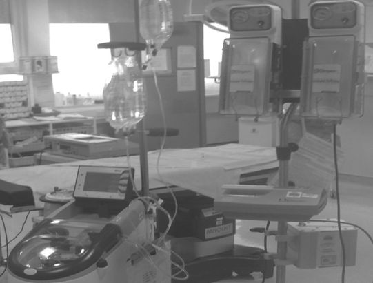
Cell-saver for ‘auto-transfusion’.
Apart from intravenous (IV) crystalloids, colloids, blood and oxytocin, the infusion of blood products needs to be considered. In massive obstetric blood loss, rapid infusion of fresh frozen plasma (FFP) may be required to replace clotting factors other than platelets. It is recommended that with every six units of blood transfusion, 1 l of FFP should be administered (15 ml/kg). Hence, 4–5 bags of FFP are required, as each bag contains about 200–250 ml of FFP. Platelet count should be maintained above 50 000 by infusing platelet concentrates, if indicated. Cryoprecipitate may also be needed if the patient develops DIC and her fibrinogen drops to less than 1 g/dl (10 g/l) [9].
Stay updated, free articles. Join our Telegram channel

Full access? Get Clinical Tree


