Posterior Mediastinal Mass
Eric J. Crotty, MD
DIFFERENTIAL DIAGNOSIS
Common
Neural Crest Tumors
Lymphoma
Less Common
Nerve Sheath Tumors
Foregut Duplication Cyst
Rare but Important
Neurenteric Cyst
Meningocele
Paraspinal Abscess
Tumor
Esophageal Varices
Extramedullary Hematopoiesis
ESSENTIAL INFORMATION
Key Differential Diagnosis Issues
Posterior mediastinal mass in young children should be considered neuroblastoma until proven otherwise
Helpful Clues for Common Diagnoses
Neural Crest Tumors
Includes neuroblastoma, ganglioneuroma, and ganglioneuroblastoma, which demonstrate spectrum of cellular maturity and malignancy
Most neuroblastoma are immature, undifferentiated, and aggressive; occurs in younger population (median age < 2 years)
Ganglioneuroma most mature and least aggressive; occurs in older population (median age ˜ 7 years)
Ganglioneuroblastoma intermediate between neuroblastoma and ganglioneuroma
Radiograph: Demonstrates elongated oval appearance with tapered borders in paraspinal location
Homogeneously solid or may contain foci of calcification, which may be fine or chunky in appearance
Separation or erosion of ribs and enlargement of neural foramina may be present
CT better demonstrates calcification (40%)
MR better demonstrates intraspinal extension
Nuclear medicine imaging with methyl-iodobenzylguanidine (MIBG) labeled with I-131 or I-123 and bone scintigraphy is mainstay of diagnosis and for monitoring response to therapy
PET/CT use is evolving and promising
Lymphoma
Mediastinum is least common site of lymphoma
Nearly always associated with sites in other mediastinal compartments
Helpful Clues for Less Common Diagnoses
Nerve Sheath Tumors
Includes neurilemoma (schwannoma), neurofibroma (plexiform and nonplexiform types), and malignant schwannoma
Sharply defined round, smooth, or lobulated paraspinal masses
Homogeneous or heterogeneous attenuation on CT with mild heterogeneous enhancement
Low to intermediate signal intensity on T1 with bright signal on T2 and mild enhancement following contrast administration
Neurofibromata may have “target” appearance on T2 and inversion recovery sequences
Higher signal peripherally and intermediate signal centrally
Neurofibromata are most commonly seen in neurofibromatosis type 1, where they are most commonly multiple
May be visible along intercostal nerves and in skin and subcutaneous tissues
Can distort ribs giving them ribbon-like appearance
Plexiform neurofibromas are more infiltrative and may extend into middle mediastinum
Foregut Duplication Cyst
Rounded fluid-filled mass with thin wall associated with esophagus
Low attenuation on CT
No enhancement following contrast administration
In general, ↓ signal on T1 and ↑ signal on T2
May have higher signal on T1 due to high protein content of fluid
In correct position, indistinguishable from bronchogenic cyst by imaging
Helpful Clues for Rare Diagnoses
Neurenteric Cyst
Contains both neural and gastrointestinal elements
Associated with vertebral anomalies
Meningocele
Herniation of leptomeninges through intervertebral foramen
May be anterior or lateral in addition to (more common) dorsal
Majority associated with neurofibromatosis, vertebral and rib anomalies
Associated with spinal anomalies; may lead to kyphosis or scoliosis
Fluid attenuation on CT
Low signal on T1 with high signal on T2 and no enhancement with contrast
Paraspinal Abscess
Discitis more common in preschool children and vertebral osteomyelitis in older children
Staphylococcus aureus most common pyogenic organism; tuberculosis most common worldwide
Discitis and osteomyelitis
Disc space narrowing with indistinct end plates
Paravertebral soft tissue mass
Tuberculosis
Disc space narrowing often later than in pyogenic osteomyelitis
Gibbus deformity
Tumor
Uncommon, except for lymphoma and neural tumors
Primary (e.g., rhabdomyosarcoma) and secondary tumors (e.g., malignant melanoma) that can occur at nearly any site in body should be considered
Esophageal Varices
Most commonly secondary to portal hypertension
Paraspinal lobulated soft tissue mass
Flow voids on MR with enhancement on MRA
Extramedullary Hematopoiesis
1 or more lobulated soft tissue masses in lower thoracic paraspinal region
Homogeneous soft tissue attenuation on CT
Homogeneous signal on MR with mild to moderate enhancement
May have expanded ribs and vertebral bodies
Image Gallery
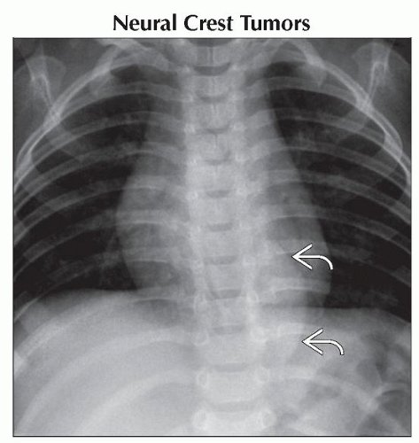 Anteroposterior radiograph shows a smoothly marginated oblong mass  in the left paraspinal location. There is no rib destruction or expansion. Calcifications are not identified. in the left paraspinal location. There is no rib destruction or expansion. Calcifications are not identified. |
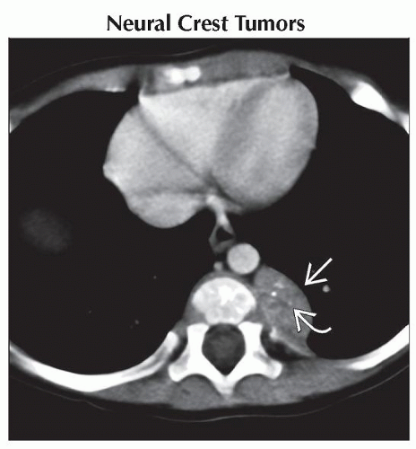 Axial CECT shows a well-circumscribed soft tissue mass  in the posterior mediastinum. There are fine calcifications in the posterior mediastinum. There are fine calcifications  within the mass, consistent with a neuroblastoma. within the mass, consistent with a neuroblastoma. |
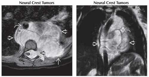 (Left) Axial T1WI C+ MR shows the mass  crossing midline and elevating the aorta crossing midline and elevating the aorta  and invading 1 of the neural foramina and invading 1 of the neural foramina  on the left. There is also evidence of invasion of the posterior thoracic wall on the left on the left. There is also evidence of invasion of the posterior thoracic wall on the left  . (Right) Coronal T2WI FSE MR in the same child shows a large mass . (Right) Coronal T2WI FSE MR in the same child shows a large mass  , predominantly within the posterior mediastinum, deviating the aorta to the right. Cystic areas of degeneration , predominantly within the posterior mediastinum, deviating the aorta to the right. Cystic areas of degeneration  are seen in this ganglioneuroblastoma. are seen in this ganglioneuroblastoma. |
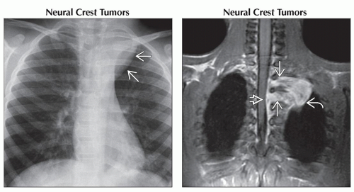 (Left) Anteroposterior radiograph shows a smoothly marginated mass
 in the superior left hemithorax. (Right) Coronal T1WI C+ FS MR shows an enhancing posterior mediastinal mass in the superior left hemithorax. (Right) Coronal T1WI C+ FS MR shows an enhancing posterior mediastinal mass  . Note the neural foramina . Note the neural foramina  with compression on the thecal sac with compression on the thecal sac  . MR is helpful in depicting neural foramina extension. Ganglioneuromas are benign tumors that may represent the end process of maturation of malignant neuroblastomas. . MR is helpful in depicting neural foramina extension. Ganglioneuromas are benign tumors that may represent the end process of maturation of malignant neuroblastomas.Stay updated, free articles. Join our Telegram channel
Full access? Get Clinical Tree
 Get Clinical Tree app for offline access

|