Chapter 404 Pleurisy, Pleural Effusions, and Empyema
Pleurisy or inflammation of the pleura is often accompanied by an effusion. The most common cause of pleural effusion in children is bacterial pneumonia (Chapter 392); heart failure (Chapter 436), rheumatologic causes, and metastatic intrathoracic malignancy are the next most common causes. A variety of other diseases account for the remaining cases, including tuberculosis (Chapter 207), lupus erythematosus (Chapter 152), aspiration pneumonitis (Chapter 389), uremia, pancreatitis, subdiaphragmatic abscess, and rheumatoid arthritis. Males and females are affected equally.
404.1 Dry or Plastic Pleurisy (Pleural Effusion)
Glenna B. Winnie and Steven V. Lossef
Etiology
Plastic pleurisy may be associated with acute bacterial or viral pulmonary infections or may develop during the course of an acute upper respiratory tract illness. The condition is also associated with tuberculosis and connective tissue diseases such as rheumatic fever.
Pathology and Pathogenesis
The process is usually limited to the visceral pleura, with small amounts of yellow serous fluid and adhesions between the pleural surfaces. In tuberculosis, the adhesions develop rapidly and the pleura are often thickened. Occasionally, fibrin deposition and adhesions are severe enough to produce a fibrothorax that markedly inhibits the excursions of the lung.
Clinical Manifestations
The primary disease often overshadows signs and symptoms. Pain, the principal symptom, is exaggerated by deep breathing, coughing, and straining. Occasionally, pleural pain is described as a dull ache, which is less likely to vary with breathing. The pain is often localized over the chest wall and is referred to the shoulder or the back. Pain with breathing is responsible for grunting and guarding of respirations, and the child often lies on the affected side in an attempt to decrease respiratory excursions. Early in the illness, a leathery, rough, inspiratory and expiratory friction rub may be audible, but it usually disappears rapidly. If the layer of exudate is thick, increased dullness to percussion and decreased breath sounds may be heard. Pleurisy may be asymptomatic. Chronic pleurisy is occasionally encountered with conditions such as atelectasis, pulmonary abscess, connective tissue diseases, and tuberculosis.
Laboratory Findings
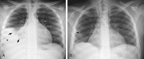
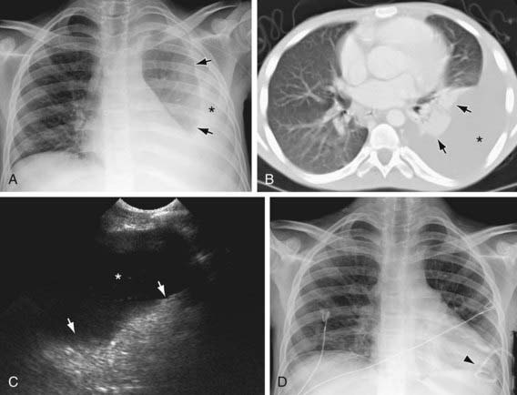
Plastic pleurisy may be detected on radiographs as a diffuse haziness at the pleural surface or a dense, sharply demarcated shadow (Figs. 404-1 and 404-2). The latter finding may be indistinguishable from small amounts of pleural exudate. Chest radiographic findings may be normal, but ultrasonography or CT findings will be positive.

Figure 404-1 A, Right pleural effusion (asterisk) due to lupus erythematosus in a 12 yr old child. Note compressed middle and lower lobes of the right lung (arrows). B, The effusion was evacuated and the right lung was completely reexpanded after insertion of the pigtail chest tube (arrow).

Figure 404-2 Left pleural effusion in a teenager with AIDS and mycoplasma avium intracellulare infection. The pleural effusion (asterisk) is clearly seen on the chest radiograph (A), CT scan (B), and ultrasonogram (C) of the left chest. Arrows point to the compressed and atelectatic left lung. D, A pigtail chest tube (arrowhead) was inserted, resulting in reexpansion of the left lung.
Differential Diagnosis
Plastic pleurisy must be distinguished from other diseases, such as epidemic pleurodynia, trauma to the rib cage (rib fracture), lesions of the dorsal root ganglia, tumors of the spinal cord, herpes zoster, gallbladder disease, and trichinosis. Even if evidence of pleural fluid is not found on physical or radiographic examination, a CT- or ultrasound-guided pleural tap in suspected cases often results in the recovery of a small amount of exudate, which when cultured may reveal the underlying bacterial cause in patients with an acute pneumonia. Patients with pleurisy and pneumonia should always be screened for tuberculosis.
Treatment
Therapy should be aimed at the underlying disease. When pneumonia is present, neither immobilization of the chest with adhesive plaster nor therapy with drugs capable of suppressing the cough reflex is indicated. If pneumonia is not present or is under good therapeutic control, strapping of the chest to restrict expansion may afford relief from pain. Analgesia with nonsteroidal anti-inflammatory agents may be helpful.
404.2 Serofibrinous or Serosanguineous Pleurisy (Pleural Effusion)
Etiology
Serofibrinous pleurisy is defined by a fibrinous exudate on the pleural surface and an exudative effusion of serous fluid into the pleural cavity. Typically it is associated with infections of the lung or with inflammatory conditions of the abdomen or mediastinum. Occasionally, it is found with connective tissue diseases such as lupus erythematosus, periarteritis, and rheumatoid, arthritis and it may be seen with primary or metastatic neoplasms of the lung, pleura, or mediastinum; tumors are commonly associated with a hemorrhagic pleurisy.
Pathogenesis
Pleural fluid originates from the capillaries of the parietal pleura and is absorbed from the pleural space via pleural stomas and the lymphatics of the parietal pleura. The rate of fluid formation is dictated by the Starling law, by which fluid movement is determined by the balance of hydrostatic and osmotic pressures in the pleural space and pulmonary capillary bed, and the permeability of the pleural membrane. Normally, only 4-12 mL of fluid is present in the pleural space, but if formation exceeds clearance, fluid accumulates. Pleural inflammation increases the permeability of the plural surface, with increased proteinaceous fluid formation; there may also be some obstruction to lymphatic absorption.
Clinical Manifestations
Because serofibrinous pleurisy is often preceded by the plastic type, early signs and symptoms may be those of plastic pleurisy. As fluid accumulates, pleuritic pain may disappear. The patient may become asymptomatic if the effusion remains small, or there may be only signs and symptoms of the underlying disease. Large fluid collections can produce cough, dyspnea, retractions, tachypnea, orthopnea, or cyanosis.

Stay updated, free articles. Join our Telegram channel

Full access? Get Clinical Tree

