Fig. 1
Distribution of hand fractures by age. Note the relative lack of bimodal age distribution previously described (From Chew 2012)
The most common phalanx fractures involve the border digits, namely, the index and small finger rays (Fig. 2). The distal phalanx is the most common location for a non-physeal injury which typically involves a crushing mechanism, and the most common location for physeal injury is the proximal phalanx. Physeal injuries are usually Salter-Harris II or III fractures. Diaphyseal fractures are more likely to occur in older populations, whereas younger patients are more likely to incur injury to the metaphysis and/or epiphysis. There is no difference in the incidence between right and left-sided hand injuries in children.
Fig. 2
Distribution of hand fractures by bone. Note the predominance of injury to the index and small finger rays (From Chew 2012)
Pathoanatomy and Applied Anatomy
The anatomy and tissue composition of the pediatric hand differ considerably from the adult. The viscoelasticity of pediatric tissue results in fracture of the bone before failure of ligaments and other soft tissues. A sound understanding of the pertinent anatomical bony and soft tissue structures is integral in order to perform an initial evaluation and develop an appropriate treatment plan for any phalanx fracture or other pediatric hand injury. One must also have an intimate understanding of both physeal and metacarpophalangeal/interphalangeal joint anatomy and the locations of attachment of soft tissues, i.e., tendons and ligaments. Particularly with distal phalanx fractures, a firm knowledge of the nail and its supporting structures is of the utmost importance.
Osseous Anatomy
Although potential epiphyses exist at both the proximal and distal ends of the metacarpal and phalanx, secondary centers of ossification develop in the proximal end of the phalanx (i.e., the base) and the distal end of the finger metacarpals (i.e., the head). This is in distinction to the thumb, in which the secondary center of ossification arises at the base of the metacarpal. Phalangeal physes in the hand remain open in boys and girls until approximately 16½ and 14½ years, respectively (Hastings and Simmons 1984). Figure 3 demonstrates the appearance of secondary ossification centers in the male and female patient, including the age at which fusion occurs to the primary centers of ossification.
Fig. 3
Appearance (A) and fusion (F) of secondary centers of ossification in the pediatric hand (From Bucholz et al. 2006)
Physeal Anatomy
The zone of hypertrophy (zone III) of the physis is an area where cellular hypertrophy results in a relative deficiency of extracellular matrix. It is the weakest layer of the growth plate, and as a result, Salter-Harris fractures tend to involve this zone. In zones I and II that abut the metaphysis, collagen provides a supportive framework, while in zone IV, provisional calcification resists pathologic forces (Torre 1988). In addition, the topography of the growth plate becomes more irregular as the patient progresses toward skeletal maturity. As a result, fractures may propagate across several zones, which may account for the partial growth arrest seen in some fractures (O’Brien 1984). However, the physis has an excellent healing potential deriving from an exuberant blood supply from both enchondral and periosteal vessels that have been shown to survive even in the event of significant displacement (Katarincic et al. 2011).
Soft Tissue Anatomy
The pediatric periosteum is a hearty structure that provides a rigid support to reduction. In addition, the periosteum delivers a healing potential as it is well vascularized and provides a population of cells that may eventually become osteoblasts when fracture healing is required. However, the thick pediatric periosteum may also become entrapped in the fracture site, resulting in an inability to obtain an adequate reduction.
The origin and insertion of tendons and ligaments are of particular importance in creating the fracture patterns commonly seen in the pediatric patient. At the level of the metacarpophalangeal joint, the collateral ligaments attach nearly entirely at the epiphyses of the adjacent metacarpal and proximal phalanx. This leads to a high incidence of Salter-Harris type II and III fractures , as the stronger collateral ligament avulses the epiphysis. The palmar plate follows a similar epiphyseal attachment pattern. In distinction, the collateral ligaments and the palmar plate of the proximal and distal interphalangeal joints have a more broad-based insertion, extending beyond the epiphysis and attaching to the metaphysis of the distal segment as well. Thus, the physis at these locations are somewhat protected from the avulsion mechanisms to which the proximal phalangeal physis is exposed (Crick et al. 1988). As a rule, the extensor tendons, including the central slip and terminal extensor tendon, insert on the epiphysis of the proximal and distal phalanges, respectively. The flexor digitorum profundus tendons and flexor pollicis longus insert on the metaphysis of the terminal phalanx, while the flexor digitorum superficialis (FDS) tendons insert over the central 60 % of the middle phalanx (Graham and Hastings 2000). Figure 4 shows the soft tissue anatomy surrounding the metacarpophalangeal (MCP) and interphalangeal joints.
Fig. 4
(a) Soft tissue attachments about the MCP joint. Note that the collateral ligaments and palmar plate insert on the epiphyseal portion of the proximal phalanx. (b) Soft tissue attachments about the proximal interphalangeal (PIP) joint. (c) Soft tissue attachments about the distal interphalangeal (DIP) joint. The collateral ligaments and palmar plates of these joints originate on the epiphysis of the proximal segment and have dual insertion on the epiphysis and metaphysis of the distal segment. The extensor apparatus attachments across the PIP joint and the DIP joint (i.e., central slip and terminal tendon) insert on the dorsal aspect of the distal epiphyseal segment, respectively. The flexor digitorum superficialis (FDS) and flexor digitorum profundus (FDP) tendons insert onto the metaphysis of the middle and distal phalanx, respectively (From Graham and Hastings 2000)
Nail Anatomy
Although nail and nail bed injuries are discussed in a separate chapter, these structures are often injured in conjunction with phalanx fractures, most commonly in association with distal phalanx or tuft fracture s. Whether the mechanism involves a crush, laceration, or avulsion injury, there can be significant damage to the nail organ. If left untreated, these injuries can result in a cosmetic deformity as well as an increased risk of infection, altered growth resulting in pain, and hygiene issues (Egol et al. 2010). Figure 5 demonstrates the distal phalanx and nail complex anatomy.
Fig. 5
Nail plate and nail bed anatomy and relationship to surrounding bony and soft tissue structures
Remodeling in Pediatric Phalanx Fractures
The pediatric patient has an incredible ability to remodel phalanx fractures as long as 2 years of growth or more remains. Angulation in the plane of joint motion (i.e., flexion-extension) is the best tolerated and can readily remodel up to 30° in children younger than 10 years of age and up to 20° after 10 years of age (Egol et al. 2010). Deformity in the coronal plane (i.e., abduction-adduction) also has the potential for remodeling, although less so than sagittal plane deformity. Residual rotational deformity following reduction has little potential for remodeling and is thus an indication for operative intervention. In addition, as fractures occur farther from the physis (i.e., more distal in the phalanx), the capacity for remodeling decreases substantially. For example, phalangeal neck fractures have a much poorer chance for obtaining acceptable alignment following completion of remodeling and healing.
Assessment
When initially evaluating a patient, the physician should also ascertain what treatments have already been initiated, including application of tourniquets in the field, providing pain medications, administration of local/systemic antibiotics including tetanus prophylaxis, and most importantly application of local, topical, or regional anesthesia which may make the physical examination difficult to interpret. Any contamination of the wounds, especially marine or barnyard contamination, should be determined. The history should also include the age, hand dominance, sport participation, and/or any avocations or jobs that the child may have. In addition, any prior injuries and their treatments or surgeries, their past medical history including allergies and medications, and their social history including who lives with them in the house should be obtained.
Physical examination of the injured child can be challenging. Children can be generally distrustful of an adult trying to touch or evaluate their painful, swollen finger. The “white coat phenomenon” can certainly play a role in the interaction between the physician and an older child or adolescent patient, and stranger anxiety or separation anxiety can provide a difficult roadblock to accurate evaluation and diagnosis. Thus, the evaluation begins upon entering the exam room by observation of the patient. The physician should be aware of the use or nonuse of certain fingers or the entire extremity. Other signs of injury potentially indicating child abuse should be investigated. These include bruising and/or fractures in various stages of healing, suspicious abrasions, or burns, among others. The interaction between the child and their parents or guardians should also be observed for warning signs of abuse. One should observe the injured digit for swelling, ecchymosis, deformity, and limited motion, all of which may be indicative of a fracture. Both the resting position of the digit and its position during active and passive motion should be examined. Residual malrotation of the digit must not be overlooked, as it is an absolute indication for operative management. Overlapping of fingers and an asymmetrical cascade compared to the contralateral hand are clues to injuries with a malrotation component. If swelling makes rotational alignment difficult to appreciate, observation of the position of the nail plates with respect to one another can provide useful information. Angular alignment is more easily appreciated.
It is crucial to assess the neurovascular status of the involved digit. This holds true for all injury mechanisms, including closed crush injuries. In terms of vascular compromise, one can look at capillary refill, differences in temperature between hands, and frank duskiness or color change of the pulp. If necessary, a Doppler exam can be performed to establish the presence of digital arterial pulses distal to the injury. Finally, pulse oximetry monitors can be applied to the fingertip and the waveforms monitored for evidence of vascular continuity.
Sensation and motor function are much harder to elicit in the young pediatric patient. Sharp versus dull and 2-point discrimination does not become a reasonable measurement of nerve function until about 5 years of age (Rang et al. 2005). In younger patients who cannot or will not verbalize sensory function, the “wrinkle test” can be employed. This test is performed by submerging the hand or digit in warm water for 5 minutes with subsequent examination of the volar finger pulp for wrinkling, indicative of retained innervation. The resting position of the digit can also provide important clues to the diagnosis, as in the case of a bony mallet finger. Passive wrist motion and observation of the tenodesis effect also provides important information. Obviously, active motor function and range of motion are a helpful adjunct in the examination. However, depending on the age and maturity of the patient, he or she may be unwilling to cooperate with this aspect of the exam. It is important to be patient and repeat the necessary components of the exam until the necessary information is obtained.
Imaging of the injured digit should begin with true anteroposterior (AP) and lateral radiographs. Oblique views can be helpful, particularly when evaluating the proximal phalanges that overlay each other on a lateral view making detailed assessment difficult. One must evaluate every digit on hand films so as not to miss injuries of adjacent digits or phalanges. It is also important to obtain a true lateral of the affected digit in isolation, particularly when intra-articular fractures are present.
Treatment of Specific Fracture Types
Distal Phalanx Fractures
Injury to the fingertip in the pediatric population occurs with great frequency, particularly during the toddler years. Associated soft tissue injuries must be identified and treated concurrently in order to optimize both functional and cosmetic outcomes. The nail and its specialized structures are particularly vulnerable in injuries of the distal phalanx. This section will provide a discussion of the following fracture types: extra-articular or tuft fractures, Seymour fractures, and bony mallet finger injuries.
Extra-articular Distal Phalanx Fractures
Distal phalanx tuft fractures are most common in toddlers due to crush injuries. Most require only symptomatic relief with analgesics, protection, and splinting for several weeks. Patients may discontinue splinting and return to activity when comfortable. The associated soft tissue and nail bed damage resulting from a fingertip crush typically becomes the focus of treatment in these injuries. An associated subungual hematoma greater than 50 % of the nail surface area should be treated with nail plate removal for hematoma evacuation and nail bed repair. Trephination with a sterile needle or pin may be attempted if the subungual hematoma involves less than 50 % of the nail bed, as this can provide improved pain relief (Fig. 6).
Fig. 6
Classification of extra-articular distal phalanx fractures. (a) Transverse diaphyseal fracture. (b) Longitudinal splitting fracture. (c) Comminuted distal tuft fracture (From Kozin and Waters 2006)
Seymour Fractures
First described by Seymour in 1966, the classic Seymour fracture is a juxtaepiphyseal fracture of the terminal phalanx with an associated laceration of the nail bed, flexion deformity, and ungual subluxation (Seymour 1966). If the physis is involved, as is typically the case in the pediatric population, usually these will be Salter-Harris type I or II fractures (Krusche-Mandl et al. 2013). The proximal edge of the nail is avulsed from the eponychial fold and usually presents as an open fracture (Barton 1979; Al-Qattan 2001a).
On physical exam, the fingertip droops and takes on the appearance of a mallet finger as a result of the flexion deformity typically produced at the fracture site. Clues as to the presence of this injury pattern include frank exposure of the proximal nail plate or in more subtle cases bleeding present near the eponychial fold. The nail bed laceration is typically not visible; however, the proximal edge of the nail plate will sit on top of the eponychial fold (Fig. 7). It is important to look for bleeding around the eponychial fold even when the proximal nail plate remains within the nail fold because the underlying nail bed can still be torn, representing an open injury.
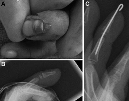
Fig. 7
Seymour fracture. (a) Clinical photo demonstrating avulsion of the proximal nail plate and bed from the eponychial fold. One must be aware that the clinical presentation and/or radiographs may be subtle. (b) In this case, radiographs demonstrate mild dorsal displacement through the distal phalanx physis. (c) As will be explained in further detail in the operative section, irrigation and debridement with K-wire fixation were eventually performed (From Yeh and Dodds 2009)
On a true lateral radiograph, there is often dorsal physeal widening. The epiphysis typically remains intact while the metaphysis is angulated. This distal segment will be flexed.
This fracture requires surgical treatment to avoid complications such as infection and growth arrest. The proximal edge of the lacerated nail matrix or nail fold is typically incarcerated in the physis. This tissue needs to be removed from the fracture site to allow fracture reduction to occur. The nail plate must be removed to allow for fracture irrigation and debridement, access to the incarcerated tissue, and soft tissue repair and/or reconstruction. For preoperative considerations, refer to Table 1 below. This checklist may be applied to the vast majority of pediatric phalanx fractures; however, any unique elements for specific fracture types will be identified.
Table 1
Preoperative considerations pertaining to the vast majority of pediatric hand fracture s
Preoperative checklist | |
|---|---|
Positioning | Patient should be positioned supine with an arm board attached to the OR table |
Fluoroscopy | Mini C-arm should be placed at the end of the arm table so that the C-arm can be placed in a perpendicular position and brought in over the arm board |
Equipment | Freer elevator, Kelly clamp, small curettes, dental pick, K-wires, K-wire driver, C-arm, chromic suture, wire cutter, heavy needle driver, arm or forearm tourniquet |
Surgical Technique
The administration of intravenous antibiotics should be performed prior to beginning the procedure. Following tourniquet inflation, the nail plate is removed. This is accomplished by first sliding a freer underneath the nail plate, then attaching a Kelly clamp, and applying a twisting/pulling motion. Once removed, the nail plate should be placed in saline and cleaned for later placement in the nail fold for fracture protection at the end of the procedure. For adequate visualization, it is often necessary to make longitudinal incisions at the proximal corners of the nail fold to allow for elevation and retraction. The laceration through the nail bed should now be visible, and the entrapped tissue may be removed.
Hyper-flexing the distal fragment will aid in exposure of the fracture site. Any gross debris and/or hematoma should be removed in the fracture site with the use of dental picks and small curettes. The site must be copiously irrigated, as this constitutes an open injury.
The fracture should be reduced and transfixed with a Kirschner (K-) wire. Depending on the size of the child, a 0.35 or 0.45 in. K-wire can be used. The K-wire can either be placed anterograde through the fracture site out the distal extent of the finger prior to fracture reduction and then passed retrograde across the proximal fragment, or the wire can be placed entirely in a retrograde fashion following fracture reduction. Whichever technique is utilized, the wire should be driven through the proximal phalanx and across the DIP joint so that it is held in extension. The wire should end in the subchondral bone of the middle phalanx. At that point, fracture reduction and pin placement are confirmed with fluoroscopy. The K-wire can either be cut beneath the level of the skin for later removal in the operating room (OR) or the wire can be left outside the skin, bent 90°, and cut.
Attention should now be turned to the nail bed laceration. If the laceration is distal to the eponychial fold, it should be repaired using 6-0 chromic suture. The longitudinal incisions at the proximal aspect of the nail fold should also be repaired using chromic suture. The nail plate that was removed at the beginning of the case should now be cleaned, trimmed if needed, and replaced beneath the eponychial fold to provide support for the fracture as well as maintain an open eponychial fold. A dressing of bacitracin, soft gauze, and Coban is applied to the digit prior to tourniquet deflation. Table 2 summarizes the key surgical steps in the treatment of Seymour fractures.
Table 2


Key steps to the surgical treatment of Seymour fractures
Stay updated, free articles. Join our Telegram channel
Full access? Get Clinical Tree
