Score as a percent of normal
Outcome tool
Joints measured
Formula for percent of normal motion
Excellent
Good
Fair
Poor
Strickland–Glogovac score
PIP and DIP
Active PIP ± DIP flexion – extension lag × 100/175°
85–100
70–84
50–69
<50
Total active motion (TAM score)
MP, PIP, and DIP
Active PIP ± MP ± DIP flexion – degrees from full × 100/260°
100
>75
>50
<50
DIP ROM score (Moiemen-Elliot 2000)
DIP
DIP range of motion × 100/74°
85–100
70–84
50–69
0–49
Table 2
Buck-Gramcko scoring system for assessing digital range of motion
Units | Points | |
|---|---|---|
Free nail palm crease distance measured from the free nail margin to the distal palmar crease | 0.0–0.5 cm | 6 |
0.6–1.5 cm | 5 | |
1.6–2.5 cm | 4 | |
2.6–4.0 cm | 3 | |
4.1–6.0 cm | 2 | |
>6.0 cm | 1 | |
Total extension deficit (MPJ + PIP + DIP) | 0°–30° | 3 |
31°–50° | 2 | |
51°–70° | 1 | |
>70° | 0 | |
Modified total active motion (MPJ + 2 × PIP + 3 × DIP) | >400° | 8 |
>320° | 6 | |
>280° | 4 | |
>240° | 2 | |
>240° | 0 | |
Classification | ||
Excellent | – | 16–17 |
Very good | – | 14–15 |
Good | – | 11–13 |
Fair | – | 7–9 |
Poor | – | 0–6 |
Flexor Tendon Treatment Options
Nonoperative Treatment
Unless the child is not medically fit for surgery, or unless the wound is too contaminated for tendon repair, most of these injuries require surgery to repair or reconstruct the tendon injury (Table 3). If a tendon is left unrepaired, not only will the digit not actively flex at the involved joints, but proximal muscle atrophy and retardation of bone growth may occur as sequelae (Bora 1970; Cunningham et al. 1985). If there is a chronic FDP injury where primary repair will not be possible and the FDS has adequate strength to flex the PIP joint, then allowing the patient to have an FDS finger is a reasonable option.
Operative Treatment
Surgery is indicated in the vast majority of flexor tendon laceration cases. If the child has incomplete flexion and pain during attempted flexion, partial laceration is possible and surgical exploration is indicated. Partial laceration of <50 % does not typically require repair, but warrants debridement of the tendon flaps to assure optimal gliding within the pulley system. Conversely, laceration of >50 % of the tendinous substance warrants surgical repair. The typical scenario in Zone II is a complete laceration of one or both of the flexor tendons. While the method of repair and the suture material chosen is universally controversial, some general themes do exist.
Preoperative Planning
In the preoperative planning stage, the surgeon must be prepared for and the patient and parents must be consented for all possibilities that could be encountered (Table 4). The tendon may or may not be amenable to primary repair, especially in cases when there is a delay from the time of injury to surgical presentation. The surgeon should account for the possibility of requiring primary tendon grafting, a two-stage tendon reconstruction, or a tendon transfer procedure. The surgeon should also plan for and obtain consent for the possibility of repair or reconstruction of a nerve or vascular structure. Silicone tendon rods, an appropriate hand holding device, suture material, microscope, and microscopic instruments should all be readily available in the operating room.
Table 3
Flexor tendon injury, nonoperative management
Flexor tendon injury nonoperative management | |
|---|---|
Indications | Contraindications |
Chronic FDP injury where FDS can flex the PIP joint | Repair can safely be performed |
Medically too ill for surgery | |
Wound too contaminated for surgery | |
Surgical Procedure, Flexor Tendon Repair
The procedure is performed under general anesthesia with the patient in the supine position. A hand table is typically used for a larger child, while an arm board or just the operating room table may be used for a smaller child. A small arm tourniquet is applied and the extremity is prepared and draped as per routine. Intravenous antibiotics should be administered within 60 min of the skin incision. Bruner incisions are preferred over midaxial incisions, especially in infants and children less than 5 years of age. Kavouksorian and Noone (1982) reported that in younger patients, midaxial incisions may migrate palmarward and result in digital flexion contractures. Both digital neurovascular bundles should be surgically explored. In contrast to the adult patient, it can be very difficult to assure the integrity of neurovascular structures based solely on the preoperative physical examination of the pediatric patient.
The tendons are subsequently explored. Windows can be made in the pulley system as necessary. The site of pulley system “windowing” should be made after careful assessment of the probable repair location. The surgeon should not open the pulley system in the same location for every case. For instance, if the tendon laceration is very distal, the A4 pulley may need to be sacrificed, but the A3 pulley may be preserved. Venting of the entire A4 pulley, or part of the A2 pulley (up to 50 %), may be required and should still allow for an acceptable result (Kwai Ben and Elliot 1998; Tang 2013).
Operative Technique
Surgical repair techniques vary depending on the author and center. The authors’ preferred suture configuration repair method for both adults and those older children with larger tendon sizes is the cross-locked cruciate–interlocking horizontal mattress (CLC-IHM) method. Both the core component of the cross-locked cruciate and the circumferential component of the interlocking horizontal mattress have superior biomechanical characteristics when compared to other commonly used methods (Figs. 1 and 2) (Croog et al. 2007; Lee et al. 2010; Lee 2012; Vigler et al. 2008). Unfortunately, there is not typically enough space in the tendon for this repair technique in small children under the age of 10 years.


Fig. 1
Cross-locked cruciate repair (Lee et al. 2010). Core sutures placed in a locked fashion prevent “trumpeting” or bulk formation at the repair site, theoretically decreasing friction and enhancing tendon gliding

Fig. 2
Interlocking horizontal mattress repair (Lee et al. 2010). Circumferential suture configuration
In children under 2 years of age, the FDP is reported to measure 2–3 mm in width and 0.5–1 mm in thickness. For this age group, most authors use a two-strand modified Kessler suture configuration (Fig. 3) (Berndtsson and Ejeskar 1995; Elhassan et al. 2006; Fitoussi et al. 1999; Grobbelaar and Hudson 1994; Herndon 1976; Kato et al. 2002; O’Connell et al. 1994). In children over 5 years of age, a four-strand repair (Fig. 4) may be feasible depending on the size of the child (Elhassan et al. 2006; Navali and Rouhani 2008; Nietosvaara et al. 2007). The rupture rates for four-strand repairs have been shown to be lower than those observed following two-strand repairs (Navali and Rouhani 2008; Nietosvaara et al. 2007).


Fig. 3
Modified Kessler repair. Suture configuration for two-strand repair technique
Al-Qattan advocates using three “figure of 8” sutures for children regardless of age. He reported higher tensile strength than the modified Kessler configuration with this technique (Al-Qattan and Al-Turaiki 2009). The chief disadvantage of the “figure of 8” repair is that the resulting repair site is bulky and oftentimes too large to freely pass underneath the flexor sheath. Therefore, venting of the flexor pulley system is almost always required. Most authors supplement the core suture with a running circumferential suture, which is still surgically practical in the pediatric patient despite the small size of the tendons. Isolated repair of the FDP, as compared to repair of both the FDP and FDS tendons, is controversial in both the adult and pediatric literature. Some authors (Al-Qattan 2013; Fitoussi et al. 1999; Kato et al. 2002) recommend only repairing the FDP tendon injury, whereas other authors (Grobbelaar and Hudson 1994) advocate repair of both. Paillard and colleagues (Paillard et al. 2002) showed that resection of one slip of the FDS tendon decreased gliding resistance as compared to pulley plasty in a cadaveric model. The subject of repair of both slips of FDS tendon, versus one slip, as compared to none is also controversial. Based on the above study and clinical experience, it is reasonable to repair one slip of FDS tendon provided that the FDP tendon still glides easily after repair. It is imperative that the tendons glide with no interruption in the operating room. It is a fallacy to think that the gliding will improve with time and motion. It is safe to conclude that the motion the surgeon achieves in the operating room is the best that will be achieved postoperatively.
In regard to the core suture purchase length, it has been shown in adults that the optimal suture purchase length biomechanically is between 7 and 10 mm (Lee et al. 2010; Tang et al. 2005). For children, Navali and Rouhani (2008) recommend the rule of a suture purchase length of 1.5–2.0 times the width of the tendon. In a series of 12 children with ages between 15 and 23 months, Al-Qattan (2013) showed that the average width of the FDP tendon in Zone II was 2.5 mm. Depending on patient size, the suture purchase length should typically equal 3.5–4.5 mm in this patient population. Navali and Rouhani (2008) also advocated that this distance should not exceed 5 mm.
Zone I Repairs
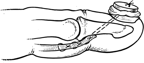
Elhassan et al. (2006) reported on the Bunnell pullout repair (Bunnell 1948) tied over a button on the nail plate (Fig. 5) in a series of 16 children. One repair ruptured postoperatively, while the remaining cases had a TAM (total active motion) of 89 % of normal which is rated as excellent (range 66–100 %). Al-Qattan (2013) advocates avoiding the possible complication of infection and nail deformity associated with the dorsal button technique by repairing the proximal tendon to the short distal stump and reinforcing it with a repair to the volar plate of the DIP at a point distal to the joint. Special care is required to avoid placing sutures deep into the physis, which may result in a growth disturbance. Al-Qattan (2012) reported on the use of this technique in a series of 10 children. There were no cases of ruptures or growth plate injury, and all patients had excellent results on the Strickland–Glogovac (1980) score. Using the Moiemen–Elliot (2000) scale, excellent results were achieved in 5 children, good results in 1, and fair results in 4.


Fig. 5
Bunnell button repair (Lee et al. 2011). A two-strand Bunnell pullout suture is passed through the distal phalanx and tied over a button on the dorsal nail plate
FPL Repair
Repair principles and outcomes are similar in the flexor pollicis longus (FPL) as compared to the FDP tendons. Grobbelaar and Hudson (1995) reported on nine children with FPL repairs managed postoperatively with controlled mobilization. Good to excellent results using Buck-Gramcko scores were achieved in seven patients, with no cases of tendon rupture reported. Fitoussi et al. (2000) reported on 16 children with FPL repairs using a modified Kessler suture repair technique. Buck-Gramcko evaluation showed good to excellent results in all but one patient who had a tendon rupture post-repair. One third of the cases had at least a 30° decrease in ROM as compared to the normal side. The authors also showed that digital nerve injury had no negative effect on eventual outcome. Overall, the authors had improved results with earlier mobilization. Orhun et al. (1999) had excellent Buck-Gramcko evaluations in 6 of 7 patients, with one remaining good result in a series of pediatric Zone II FPL repairs. In the setting of a chronic FPL injury, good results have been reported with a two-stage tendon transfer utilizing a silicone tendon implant followed by a transfer of the FDS of the ring finger (Yamamoto and Fujita 2013).
Preferred Methods: Tendon Repair
The authors’ preferred methods for managing Zone II flexor tendon injuries by age are shown in Tables 5, 6, and 7. In all age groups, Bruner incisions are utilized, the location of tendon repair is identified, and appropriate windows are made in the pulley system for access. In children <5 years, a modified Kessler (Fig. 3) suture with 4-0 Fiberwire (Arthrex, Naples, FL, USA) is performed, with a purchase length determined by tendon size. A circumferential suture with Prolene (Ethicon, Somerset, NJ, USA) is then placed in an interlocking horizontal mattress fashion (Fig. 2). If the FDS tendons are involved, repair of one slip is performed in a “figure of 8” fashion. Venting of the pulleys is performed where necessary to optimize gliding. The core suture technique is dependent upon the patient’s age and anatomy. In children 6–10 years of age, a Strickland repair (Fig. 4) is performed as permitted by the flexor tendon size, and core suture purchase length is adjusted to tendon size. In older children (>10 years), a cross-locked cruciate core suture is preferred as size permits.
Table 4
Preoperative planning for pediatric flexor tendon repair
Flexor tendon injuries – preoperative planning |
|---|
OR table: hand table >10 years, arm board 5–10 years, OR table <5 years |
Position/positioning aids: supine |
Equipment: hand holder, tendon passer, microscope, and instruments |
Tourniquet |
Consented for tendon graft, silicone rod, tendon transfer |
Consented for nerve and artery repair versus reconstruction |
Table 5
Preferred management of Zone II injuries in children under 5 years
Flexor tendon injuries – surgical steps, Zone II <5 years |
|---|
Bruner incisions |
Identify location of tendon repair |
Open appropriate area of pulleys |
Avoid handling tendon surface |
Hold tendon ends with 25 gauge needles |
For <5 years, modified Kessler (Fig. 3) with 4-0 Fiberwire |
Suture purchase of core suture: 4 mm |
Interlocking horizontal mattress circumferential suture (Fig. 2) with 6-0 or 7-0 Prolene |
Circumferential suture with 6-0 or 7-0 Prolene |
Repair 1 slip of FDS with figure of 8 suture, 4-0 to 6-0 Prolene |
Vent pulleys as necessary |
Table 6
Preferred management of Zone II injuries in children under 6–10 years
Flexor tendon injuries – surgical steps, Zone II, 6–10 years |
|---|
Bruner incisions |
Identify location of tendon repair |
Open appropriate area of pulleys |
Avoid handling tendon surface |
Hold tendon ends with 25 gauge needles |
Modified Kessler core suture with 4-0 Fiberwire |
Possibly Strickland repair (Fig. 4) if anatomy permits |
Suture purchase of core suture: 4–5 mm |
Interlocking horizontal mattress suture with 6-0 Prolene |
Repair 1 slip of FDS with figure of 8 suture, 4-0 to 6-0 Prolene |
Vent pulleys as necessary |
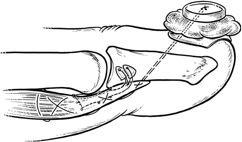
The authors’ preferred technique for managing Zone I injuries is shown in Table 8. A Bunnell stitch is performed with 2-0 to 4-0 Prolene depending on the tendon size, which is passed with a Keith needle through the distal phalanx distal to the physis and tied over a button on the nail plate (Fig. 5). Excessive pressure on the nail plate is avoided, and the distal pulley system is vented as needed to optimize gliding. If the patient is over 10 years of age and the tendon size permits, a volarly placed pullout suture is augmented with a second suture on the dorsal aspect of the tendon that is anchored to distal phalanx with two suture anchors (Mitek Microfix Quickanchors; DePuy Mitek, Inc., Raynham, MA). This anchor-button technique (Fig. 6) has been shown to closely approximate native FDP tendon–bone interface ultimate strength, with a mean load to failure of 115 Newtons in a cadaveric model (Lee et al. 2011).

Table 7
Preferred management of Zone II injuries in children >10 years (depending on size of patient)
Flexor tendon injuries – surgical steps, Zone II, >10 years |
|---|
Bruner incisions |
Identify location of tendon repair |
Open appropriate area of pulleys |
Avoid handling tendon surface |
Hold tendon ends with 25 gauge needles |
Cross-locked cruciate (CLC) core suture (Fig. 1) with 3-0 to 4-0 Fiberwire |
Modified Kessler (MK) and possibly Strickland if size precludes CLC |
Suture purchase of core suture: 6+ mm (up to 10 mm if adult size) |
Interlocking horizontal mattress circumferential suture |
Circumferential suture with 6-0 Prolene |
Repair 1 slip of FDS with figure of 8 suture, 4-0 to 6-0 Prolene |
Vent pulleys as necessary |

Fig. 6
Anchor-button repair (Lee et al. 2011). A two-part repair consisting of a two-strand locking Krackow suture along the dorsal aspect of the tendon that is fixed with two retrograde anchors. This is supplemented with a two-strand Bunnell suture along the volar aspect of the tendon that is tied over a dorsal button
Table 8
Preferred management of Zone I injuries in pediatric patients
Flexor tendon injuries – surgical steps, Zone I
Stay updated, free articles. Join our Telegram channel
Full access? Get Clinical Tree
 Get Clinical Tree app for offline access

|
|---|
