Pediatric and Adolescent Gynecology


Pediatric and Adolescent Gynecology


Mariel A. Focseneanu

Diane F. Merritt


Pediatric and adolescent gynecology is a distinct subspecialty involving the unique gynecologic problems of the neonate, prepubertal child, and developing adolescent. A practicing obstetrician-gynecologist should be able to recognize, treat, or triage the most common officebased problems that arise in this discipline and know when to refer to a specialist.


NORMAL FINDINGS

Training in obstetrics and gynecology focuses on the care and management of the expectant and puerperal mother and much less routinely on the examination of a newborn after delivery. However, it is good practice for the delivering clinician to perform a brief genital examination of every newborn to determine if the external genital structures, urethra, and anus appear to be normal. The breast tissue of newborns of both genders may have breast buds or secretions due to maternal estrogen. In the newborn female, presence of a patent vagina must also be assessed. The female infant initially has prominent labia and a thickened hymen. Vulvar edema and a mucous vaginal discharge are common. If a hymenal polyp or vaginal “tag” is noted on the newborn examination, conservative management is recommended as spontaneous involution often occurs as endogenous estrogen levels fall. Approximately 10% of newborns will have some withdrawal bleeding. In premature infants, relative clitoromegaly may appear as the result of decreased subcutaneous fat. These expected physiologic effects of maternal hormones usually resolve within the first 3 months of life and are not a cause for concern.1

Once newborn levels of estrogen decline, the hymenal membrane thins and may become translucent allowing fine vascular details to be seen, and the introital mucosa appears shiny and reddened. The labia minora are small structures usually measuring less than half the length of the introitus, reflecting the lack of endogenous estrogen production. Prepubertal children, lacking lactobacilli, will normally have minimal clear, alkaline vaginal discharge. There are several normal variants of hymen configuration, for example, annular (circumferential), crescentic, and fimbriated (folded). The crescentic hymen was most commonly found among girls surveyed at age 3 years, with an annular hymen being the second most common configuration.2

In a prepubertal female, the size ratio of the uterine corpus compared with the cervix is 1:3; in the pubertal child, it is 1:1; and in the adult woman, it is 3:1. The prepubertal ovary is generally about 1 cm3 in size, and small follicles are seen by high-resolution ultrasound.3


Assessment of External Genitalia and Exam Techniques

The American College of Obstetricians and Gynecologists recommends the first visit to the obstetrician-gynecologist for screening and the provision of reproductive preventive health care services and guidance take place between the ages of 13 and 15 years.4 The American Academy of Pediatrics promotes the inclusion of the gynecologic examination in the primary care setting within the medical home.5 For this reason, providers who see or treat children or adolescents should become familiar with providing gynecologic examinations for infants, children, and adolescents.


Newborn and Prepubertal Child

When examining a young child, it is important to put the child and parents at ease and gain their trust. The physician should explain to the child and her parent(s) exactly what will be done during the exam and why it is necessary. Focus should be placed on reassuring the parent(s) and explaining that the examination of a child is different from an examination of an adult. The child should be made to feel at ease and allowed to maintain some control of her environment; the goal is to examine the child without causing any traumatization to her. It may be helpful to allow the child to hold a handheld mirror so she may see her genitals at the same time they are being examined.

Children and younger adolescents are concrete thinkers—as such the most meaningful information may be imparted concurrent with the examination. It is important to point out to children after the exam that it was done because the parent or guardian agreed to it. This provides an opportunity to remind children that no one else should touch her genitals and if it happens, she should tell the parent(s) or guardian(s).

The genital examination should be incorporated into a complete physical examination. Begin the examination by visual inspection of the skin for rashes and lesions, and then listen to the heart and lungs. Visualization or palpation of the breast tissue allows determination of Tanner staging. The abdomen should be palpated for masses. The pelvic exam may be deferred until a subsequent visit in order to allow the girl time to develop a relationship with her new physician or provider unless there is a specific problem or urgent situation.

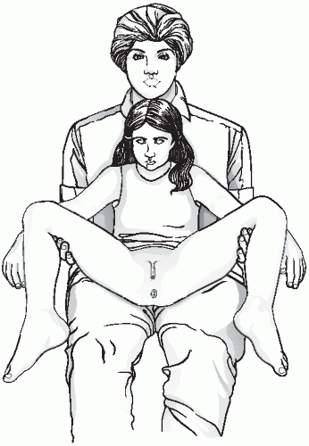
The undergarments may be removed immediately before the examination and the child should be offered and shown how to use a drape. Although the ideal position for good visualization of a young girls genitalia is supine, with her buttocks at the end of a gynecologic examination table, this is not typically feasible in younger children. Alternatively, the parent of a child can be positioned on the examination table, supported by elevating the head of the table, with the child positioned frog-leg style on the parent’s lap (
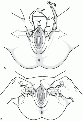
Fig. 23.1). Many children feel comforted in the arms and lap of their parent and it is an easy alternative method to allow inspection of the perineum. In the frog-leg position, the labia may be gently separated or gentle lateral traction on the labia majora may be used and this allows for better visualization of the hymen and vaginal orifice (Fig. 23.2A,B). The child may also be allowed to use her own hands to spread the labia laterally to allow visualization of the vaginal introits. After examination in the frog-leg style, the child should be asked to get into the knee-to-chest position on the examination table (Fig. 23.3). The kneeto-chest position offers an improved view of the vagina and more thorough inspection of both the posterior and anterior hymenal area.

Older patients may be able to be placed in the supine position but this position does not allow direct eye contact between the patient and her examiner. For this reason, we suggest that older children and teens sit up with their back supported at a 45-degree angle and their feet in stirrups. With the perineum exposed, a teaching examination can proceed (see next section).


FIGURE 23.1 Picture of child in frog leg in parent’s lap. (Used with permission from Finkel MA, Giardino AP, eds. Medical Evaluation of Child Sexual Abuse: A Practical Guide. 2nd ed. Thousand Oaks, CA: Sage Publications; 2002. © Sage Publications, Inc.)

Examination of a prepubertal child should not be done without some means of magnifying the area. A handheld magnifying glass with or without light, colposcope, or even an otoscope with the truncated ear canal piece removed may be used. The exam should be described in detail in the chart with a comment regarding the size, shape, or any abnormalities of the labia majora and minora (in children it may be hard to distinguish between these), clitoris, clitoral hood, urethra, vaginal orifice and vestibule, hymen, perineal body, and anus inclusive.

Because of their hypoestrogenic state, the genital tissues of young girls are very sensitive to touch and may be easily lacerated by instruments and even cotton tip swabs. A vaginal speculum should not be used in the pediatric patient. If a vaginal infection needs to be ruled out, a culture should be obtained by placing a very small moistened cotton swab, for example, a Calgi swab or a
small Dacron swab (male urethral size), just through the hymen with every effort made not to touch the sensitive hymen. If this is not possible, the culture swab may be placed between the labia, although this may not yield adequate sampling for diagnosis.


FIGURE 23.2 A, B: Separation and lateral traction. (Used with permission from Finkel MA, Giardino AP, eds. Medical Evaluation of Child Sexual Abuse: A Practical Guide. 2nd ed. Thousand Oaks, CA: Sage Publications; 2002. © Sage Publications, Inc.)

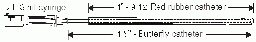
If the vagina needs to be flushed, for example, looking for a foreign body, a soft rubber catheter works quite well. However, it is not ideal for obtaining secretions for culture because the tip will be sucked against the vaginal wall when the vaginal fluid (either naturally occurring or the combination of saline that has been introduced into the vagina via the catheter and then is aspirated) is aspirated. One option is to use a “catheter within a catheter” (Fig. 23.4). A 4.5 in butterfly intravenous (IV) tube may be passed into the distal 4.5 inches of a soft, no. 12, red rubber catheter that is attached to a 1 to 3 mL syringe to allow for 0.5 to 1 mL of fluid to be flushed into the vagina and aspirated back into the syringe. This can be done several times to get a good mixture of upper vaginal secretions and the aspirant may be sent for wet mount, Gram stains, cultures, and forensic material. The lab should be told that the specimen was obtained from a prepubertal child. Most children are amenable to this procedure if they are shown there is no needle attached to the apparatus and it may be helpful to place a few drops of the liquid on their hand or fingers to reassure them.


FIGURE 23.3 Child in the knee-to-chest position. (From Gibbs RS, Karlan BY, Haney AF, Nygaard IE, eds. Danforth’s Obstetrics and Gynecology. 10th ed. Philadelphia: Lippincott Williams and Wilkins; 2008.)


FIGURE 23.4 Catheter within a catheter. (Used with permission from Pokorny SF, Stormer, J. A traumatic removal of secretions from the prepubertal vagina. Am J Obstet Gynecol.1987;156:S81-S82. © 1987 Mosby.)

The vaginal pH in prepubertal girls is 6.5 to 7.5 and is therefore not useful for the diagnosis of bacterial vaginosis or trichomonal infections in these girls.

If a more thorough examination of the vagina of a prepubertal child is necessary (e.g., unexplained vaginal bleeding, suspicion of a foreign object or trauma), vaginoscopy is recommended. The vaginal endoscopic examination via cystoscopy or hysteroscopy employs normal saline (in a bag draining to gravity) to distend the vagina while the labia are held gently together, allowing the vagina to distend and facilitate evaluation. Application of 2% lidocaine jelly to the external vaginal area and to the tip of a vaginoscope enables more comfortable placement into the vagina. This may be accomplished in a cooperative patient in an outpatient setting or may require general anesthesia in a young or frightened child.6 A young child’s vagina is about 5 cm in length and the mucosa is red, thin, and slightly folded. The cervix is small, with a central opening, and is often flush with the vagina. Vaginoscopy may cause petechiae of the vaginal mucosa given how thin and sensitive it is.

Clitoral size may be determined by the clitoral index, which is calculated as the product of the length and width of the clitoris and expressed in square millimeters. Charts indicating the normal clitoral index by age are available (Table 23.1). If the clitoris is enlarged, causes of androgen stimulation should be sought (Table 23.2).

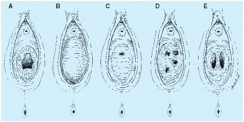
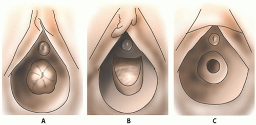
It is important to realize that hymens change with age and weight. Infant girls have estrogenized hymens and
may have a prominent ridge at 6 o’clock. The hymen of prepubertal girls is unestrogenized, thin, and may have a multitude of appearances (Figs. 23.5 and 23.6).


TABLE 23.1 Clitoral Index in Girls With Normal and Abnormal Sexual Development


Age (yr)


Normal (n)


Congenital Adrenal Hyperplasia (n)


Central Precocious Puberty (n)


Premature Adrenarche (n)


XO/XY karyotype (n)


0-1


15.1 ± 1.4 (16)


137.9 ± 26.9 (6)


1-8


15.1 ± 0.9 (29)


225.0 ± 49.0 (5)


20 ± 2 (5)


26 ± 1 (4)


8-13


16.7 ± 0.9 (18)


212.0 ± 59.0 (4)


17 ± 2 (5)


25 ± 7 (4)


242 ± 171 (2)


13-18


20.7 ± 1.6 (17)


116.0 ± 14.0 (5)


All values are clitoral indexes in square millimeters.


From Sane K, Pescovitz OH. The clitoral index: a determination of clitoral size in normal girls and in girls with abnormal sexual development. J Pediatr. 1992;120(2, pt 1):264—266.


TABLE 23.2 Causes of Clitoromegaly


Can Present


Condition


At Birth


After Birth


Comments and Reference


Virilized XX


Congenital adrenal hyperplasia (CAH)


Yes


Yes


21-Alpha-hydroxylase deficiency and 11-beta-hydroxylase deficiency are the most common. Late presentation does occur.a,b (See “Diagnosis of classic congenital adrenal hyperplasia due to 21-hydroxylase deficiency” and see “Congenital adrenal hyperplasia due to 11-beta-hydroxylase deficiency”).


Maternal androgens or synthetic progestational agents


Yes


No


(See “Causes of gestational hyperandrogenism”)


Placental aromatase enzyme deficiency


Yes


No


Maternal virilization during pregnancy can be a clue to the diagnosis. (See “Diagnosis and management of gestational hyperandrogenism”).


Androgen-secreting tumors


No


Yes


Ovarianc and adrenald


Hepatic dysfunction


No


Yes


Report of 10-year-old girl with portal hypertension and impaired hepatic steroid metabolisme


Exogenous androgens


Yes


Yes


(Akcam, 2003)f


Feminized XY (not true clitoromegaly, but small penis can appear as large clitoris)


Congenital adrenal hyperplasia (CAH)


Yes


Yes


Examples include 17-hydroxylase deficiency, 3-beta-hydroxysteroid deficiency. (See “Uncommon causes of congenital adrenal hyperplasia”).


Androgen insensitivity syndrome (aka testicular feminization syndrome)


Yes


Yes


Complete insensitivity usually presents at birth, whereas partial insensitivity can present at birth or later.g,h (See “Diagnosis and treatment of disorders of the androgen receptor”).


5-Alpha-reductase deficiency (5aRD)


Yes


No


5aR converts testosterone to DHT. (See “Steroid 5-alpha-reductase 2 deficiency”).


Other


True hermaphroditism


Yes


Yes


Patients can be XX or XY. Genitalia can vary from ambiguous to isolated clitoromegaly.i


Mixed gonadal dysgenesis


Yes


No


Most patients are mosaics 45,XO/46,XY. (See “Evaluation of the infant with ambiguous genitalia”).


Neurofibromatosis


Yes


Yes


Has been reported both with type 1 and type 2j


Vascular malformation


TP


Yes


Report of 5-year-old girlk


Sebaceous cyst


TP


Yes


Case report in adolescent girll


Idiopathic


Yes


Yes


Report of two adults with acquired isolated clitoromegalym


TP, theoretically possible but no case report found.


a Silverman K, Couey SM, Murmam D. Non-classic 21-hydroxylase deficiency in an 18-year-old female athlete. A case report. J Pediatr Adolesc Gynecol. 2000;13:96-97.

b Pang S. Congenital adrenal hyperplasia. Baillieres Clin Obstet Gynecol. 1997;11:281-306.

c Sayer RA, Deutsch A, Hoffman MS. Clitoroplasty. Obstet Gynecol. 2007;110:523-525.

d Wolthers OD, Cameron FJ, Scheimberg I, et al. Androgen secreting adrenocortical tumors. Arch Dis Child. 1999;80:46-50.

e Speiser PW, Susin M, Sassano H, et al. Ovarian hyperthecosis in the setting of portal hypertension. J Clin Endocrinol Metab. 2000;85:873-877.

f Akcam M, Topaloglu A. Extremely immature infant who developed clitoromegaly during the second month of her postnatal life probably due to frequent whole blood transfusion from an adult male. Pediatr Int. 2003;45:347-348.

g Sultan C, Lumbroso S, Paris F, et al. Disorders of androgen action. Semin Reprod Med. 2002;20:217-227.

h Chipashvili MK, Kristesashvili DI, Kopaliani NSh. Androgen insensitivity syndrome in adolescents [in Russian]. Georgian Med News. 2006;131:21-24.

i Yordam N, Alikasifoglu A, Kandemir N, et al. True hermaphroditism: clinical features, genetic variants and gonadal histology. J Pediatr Endocrinol Metab. 2001;4:421-427.

j Yuksel H, Odabasi AR, Kafkas S, et al. Clitoromegaly in type 2 neurofibromatosis: a case report and review of the literature. Eur J Gynaecol Oncol. 2003;24:447-451.

k Haritharan T, Islah M, Zulfiqar A, et al. Solitary vascular malformation of the clitoris. Med J Malaysia. 2006;61:258-259.

l Guelinckx PJ, Sinsel NK. An unusual case of clitoral enlargement. Acta Chir Belg. 2002;102:192-195.

m Copcu E, Aktas A, Sivrioglu N, et al. Idiopathic isolated clitoromegaly: a report of two cases. Reprod Health. 2004;1:4.


Data from Drutz JE. The pediatric physical examination: The perineum. Up To Date Web site. http://www.uptodate.com/contents/the-pediatric-physical-examination-the-perineum. Accessed December 17, 2013.


FIGURE 23.5 Anatomic variations of the normal hymen. A: Normal. B: Imperforate. C: Microperforate. D: Cribriform. E: Septate. (Reproduced with permission from Laufer MR. Structural abnormalities of the female reproductive tract. In: Emans SJ, Laufer MR, Goldstein DP, eds. Pediatric and Adolescent Gynecology. 6th ed. Philadelphia: Lippincott Williams & Wilkins; 2012. Copyright © 2012 Lippincott Williams & Wilkins.)

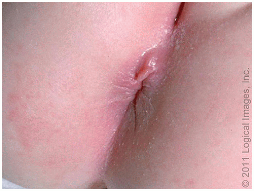
Infantile perianal pyramidal protrusion (IPPP) is a perianal lesion that has been previously described as a skin tag or fold and is seen almost only in girls7 (Fig. 23.7). It may be perineal in location rather than perianal but is typically midline and usually anterior to the anus, although they may rarely be posteriorly located to the anus.8 It is not tender and treatment is not indicated. Clinically, they may undergo cycles of swelling followed by spontaneous resolution.


Pubertal and Postpubertal Adolescent

In the initial examination of the young adolescent, often only the external genital area needs to be viewed, in which case placement of a speculum or an internal examination is not necessary. This often reassures both the patient and her parent who may have negative attitudes about the use of a vaginal speculum and allows the clinician to present the external genital examination in the context of routine adolescent health care.


FIGURE 23.6 Normal hymens in prepubertal girls. A: Fimbriated or redundant hymen. B: Posterior rim or crescentic hymen. C: Circumferential or annular hymen. (Modified from Pokorny SF. Configuration of the prepubertal hymen. Am J Obstet Gynecol. 1987;157:950.)

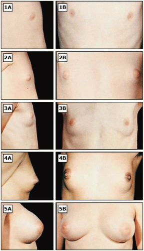
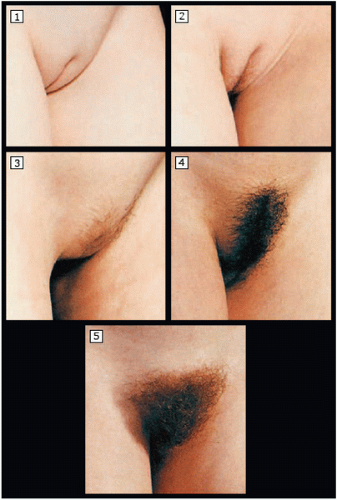
It is important for clinicians to be familiar with normal variations of pubertal maturation, so that abnormalities of puberty can be recognized and treated in a timely manner. Puberty consists of a series of predictable sequence of events and the most common staging system is the “Tanner stages” of puberty (Figs. 23.8, 2.9 to 23.10).

Normal puberty begins when gonadotropin-releasing hormone (GnRH) is released from the hypothalamus in a pulsatile manner, activating the release of luteinizing hormone (LH) and follicle-stimulating hormone (FSH)
from the pituitary. LH induces production of androstenedione from the ovarian theca cells, and FSH induces synthesis of estradiol from the follicular cells. At puberty, changes related to estrogen include breast development, enlargement of the labia minora, and edematous and pink changes in the hymenal tissues. The increase in estradiol causes breast development (thelarche) and the growth spurt in girls.9 The earliest secondary sexual characteristic in most girls is breast/areolar development, although some may exhibit pubic hair growth first. Menarche occurs, on average, 2.6 years after the onset of puberty.


FIGURE 23.7 Infantile perianal pyramidal protrusion. Infantile perianal pyramidal protrusion is a pedunculated perineal lesion, typically located in the midline, just anterior to the anus. It occurs almost exclusively in girls. (Reproduced with permission from www.visualdx.com. Copyright © 2011 Logical Images, Inc.)

Adrenarche (pubic hair development) is initiated by adrenal androgen production and usually begins approximately 6 months after thelarche, at an average age of 11 to 12 years. Pubic hair may be a normal first sign of development in some girls (as early as age 7 years), particularly in those of African American descent.10 The Lawson Wilkins Pediatric Endocrine Society has recommended guidelines for the valuation of premature development, which state that pubic hair or breast development requires evaluation only when it occurs before age 7 years in non-African American girls and before age 6 years in African American girls.11 These recommendations are controversial, however, as they lower the age threshold for evaluation for precocious puberty and this may lead to failure to identify some children with disease processes that may be amenable to intervention.12

Menarche typically occurs within 2 to 3 years after thelarche, at a median age of 12.4 years.13 Onset of puberty is affected by race, family history, birth weight, nutrition, international adoption, and exposure to estrogenic chemicals. For reasons that remain unclear, the age of menarche has declined to a greater extent in Black girls compared to White girls over the last several decades.14 When menarche occurs before the age of 12 years, it is associated with increases in weight and body mass index (BMI).
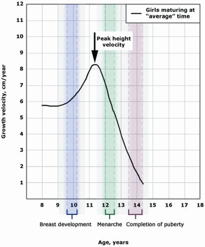
15

About 17 to 18% of final adult height accrues during puberty, with the limbs exhibiting an accelerated growth prior to the truncal portions of the body.16 The peak growth velocity usually begins about 2 years earlier in girls than in boys and occurs about 6 months or so prior to menarche.17 About half of a woman’s total body calcium is laid down during puberty.18,19 Changes in bone growth and mineralization patterns may put some adolescents at increased risk for fracture. If a patient has scoliosis, progression of the degree of scoliosis may occur during puberty as a result of growth in the axial skeleton.

The increase in BMI prior to 16 years of age that is seen with puberty is usually due in increases in fat-free mass; after age 16 years, increasing BMI is primarily due to increases in fat mass.20

The vagina and hymen become more distensible during puberty. Despite historical documentation and cultural beliefs, recent research has shown that even an experienced clinician cannot tell if a postpubertal woman has had genital penetration based on the physical examination.21

Any signs of inflammation, lesions or pigment changes seen on the exam should be noted. Folliculitis, presenting as tender papules and pustules, has become increasingly common in adolescents who shave their pubic hair.5 As noted earlier, an enlarged clitoris (>10 mm3) may indicate elevated androgens. Asymmetry of the labia, where the right and left labia are different in size and appearance, is a normal variant. Some young women are uncomfortable with asymmetric labia or enlarged labia minora and complain about self-consciousness and discomfort while wearing clothing, exercising, or having intercourse. The American College of Obstetricians and Gynecologists (ACOG) does not support performing cosmetic surgical procedures of labia minora unless there is significant impairment in function.22

A physiologic clear vaginal secretion may be worrisome for a peripubertal girl or young teen. Teens should be reassured that this nonirritating, nonodorous discharge is normal physiologic leukorrhea and reflects the effect of estrogen on the cells lining the vagina. Peri- and postpubertal children will have normal vaginal secretions with an acidic pH due to the effect of the protective population of lactobacilli that thrive in the glucose-rich environment of an estrogenized vagina.

The examination of adolescents is a golden opportunity to teach genital anatomy and hygiene. Teens appreciate a nonjudgmental and unhurried provider who is willing to answer their questions. As in the pediatric
patient, examination of the adolescent breast should include Tanner staging. Self-examination of the breast for those aged 13 to 18 years is not recommended because the risk for breast pathology is low.23 The pelvic exam should be explained prior to beginning the exam. In general, all adolescents benefit from an educational external genital examination, with the provider indicating the anatomic findings while the patient simultaneously watches using a handheld mirror. Instruction in genital hygiene, including wiping from front to back; using wet wipes after bowel movements; cleansing the folds of the labia majora and minora during baths or showers; and avoidance of douching, chemical irritants, and perfumed or deodorant soaps is crucial advice for young women.


FIGURE 23.8 Sequence of puberty in girls. Sequence of events in girls with average timing of pubertal development in the United States. Black girls tend to reach a milestone at a younger age (left-hand side of the bracket) than White girls (right-hand side of the bracket). The median length of time between the onset of puberty (breast Tanner stage II) and menarche is 2.6 years, and the 95th percentile is 4.5 years. (Data from Biro FM, Huang B, Crawford PB, et al. Pubertal correlates in black and white girls. J Pediatr. 2006;148:234-240; Tanner JM, Davies PS. Clinical longitudinal standards for height and height velocity for North American children. J Pediatr. 1985;107:317-329; Biro FM. Normal puberty. UpToDate Web site. http://www.uptodate.com/contents/normal-puberty. Accessed December 17, 2013.)

If testing for sexually transmitted infections (STIs) (either due to abuse or sexual activity) is indicated, a speculum examination is not required to obtain an endocervical specimen given the current availability and efficacy of urine-based gonorrhea and chlamydia nucleic acid amplification testing (NAAT).24 The recently revised 2009 ACOG guidelines for cervical cancer screening state that the first Pap test should be performed at age 21 years regardless of the age of first intercourse.25 The exception to this general rule is if the patient has HIV and is sexually active, initial screening should include a Pap test and pelvic exam; the Pap test should be obtained twice during the first year after diagnosis of HIV infection and, if the results are normal, annually thereafter.26

If necessary to evaluate abnormal bleeding or other pathology in a young teen, a narrow Pederson speculum may be used to visualize the cervix and vaginal mucosa. To assess a possible pelvic mass, a young adolescent, even one who has never been sexually active, may undergo a single digit bimanual exam or rectal exam.
Alternatively, a transabdominal ultrasound may be used to document internal genital structures as long as the bladder is well-filled.


FIGURE 23.9 Sexual maturity rating (Tanner staging) of breast development in girls. Stages in breast development in girls. Stage 1: Prepubertal, with no palpable breast tissue. Stage 2: Development of a breast bud, with elevation of the papilla and enlargement of the areolar diameter. Stage 3: Enlargement of the breast, without separation of areolar contour from the breast. Stage 4: The areola and papilla project above the breast, forming a secondary mound. Stage 5: Recession of the areola to match the contour of the breast; the papilla projects beyond the contour of the areola and breast. (Reproduced from Roede MJ, van Wieringen JC. Growth diagrams 1980: Netherlands third nation-wide survey. Tijdschr Soc Gezondheids. 1985;63:1, with permission.)

Anemia and iron deficiency are common among adolescent girls, reflecting the changes in hemoglobin and serum ferritin concentrations, which are influenced by both sex and pubertal stage. Both hemoglobin and serum ferritin concentrations increase in adolescent boys but not in girls; coupled with menstrual bleeding and insufficient anemia, this predisposes pubertal girls to anemia.27


VULVAR DISORDERS


Vulvovaginitis

Vulvovaginitis is the most common indication for referral to a pediatric gynecologist, is the most common gynecologic problem in prepubescent girls, and is often the result of poor hygiene or chemical irritants in combination with prepubertal vulvar anatomy.28 Prepubertal genital tissues are vulnerable to infection and irritation because there are no protective lactobacilli, the alkaline state of the vagina (pH 7), poor labial development, and the tissues are thin due to the low estrogen state. Additionally, the distance from the anus to the vestibule is comparatively short, and fecal pathogens easily access the vagina.

Symptoms and signs of vulvitis include pruritus, tenderness, dysuria, and erythema of the vulva. The presence of discharge is more indicative of a vaginitis, which is inflammation of the vagina. These two diagnoses may occur simultaneously. The differential diagnosis of vulvovaginitis in children and adolescents includes infections, vaginal foreign bodies, pinworms, trauma, lichen sclerosus, psoriasis, eczema, contact dermatitis, scabies, ectopic ureter, sexual abuse, congenital abnormalities, chronic masturbation, and commonly, poor genital hygiene. Nonspecific vulvovaginitis is vulvovaginal irritation without an identifiable bacterial pathogen, and it accounts for 25 to 75% of all cases.29 Parents and children need to understand the importance of proper hygiene in the prevention of vulvovaginitis. Carefully washing between labial folds with a nonirritating soap may prevent complaints of pain or itching in the genital region. The child should avoid soaking in a tub with shampoo or bubble bath because these may cause chemical irritation. However, soaking in warm clean bathwater for 15-minute intervals is soothing and helps with cleaning the area. We emphasize wiping from front to back (from urethra toward anus) to minimize contamination of the vulva and vagina with enteric organisms, and encourage the use of a wet wipe (front to back) after bowel movements for girls who have difficulty with recurrent vulvovaginitis. Time spent in tight-fitting clothing such as tights, leotards, and swimsuits should be minimized; loose-fitting cotton undergarments and clothing are preferred. Sleeping in nightgowns without underwear in lieu of pants or sleeper pajamas are preferable because they allow the air to circulate. Undergarments should be washed with a mild, unscented detergent, and fabric softeners should not be used. Thong underwear can promote fecal contamination of the vagina and should be discouraged. Spending prolonged periods of time in wet bathing suits should be avoided.


FIGURE 23.10 Sexual maturity rating (Tanner staging) of pubic hair development in girls. Stages of development in pubic hair in girls. Stage 1: Prepubertal with no pubic hair. Stage 2: Sparse, straight hair along the lateral vulva. Stage 3: Hair is darker, coarser, and curlier, extending over the mid-pubis. Stage 4: Hair is adult-like in appearance but does not extend to the thighs. Stage 5: Hair is adult in appearance, extending from thigh to thigh. (Reproduced from Roede MJ, van Wieringen JC. Growth diagrams 1980: Netherlands third nation-wide survey. Tijdschr Soc Gezondheids. 1985;63:1, with permission.)


In a pediatric population, infectious vulvovaginitis, where a specific pathogen is isolated as the cause of symptoms, may be caused by fecal or respiratory pathogens. Cultures most commonly reveal Escherichia coli; Group A beta-hemolytic Streptococci (Streptococcus pyogenes) or Haemophilus influenzae. Other respiratory pathogens include Staphylococcus aureus, Streptococcus pneumoniae, Branhamella catarrhalis, and Neisseria meningitidis. Other fecal organisms that may be cultured include Shigella and Yersinia. Streptococcus pyogenes is the most commonly identified pathogen in prepubertal girls (up to 20%).30 These organisms are generally transmitted by hand to the genital area as a result of poor hand washing or improper toilet hygiene. A specific antimicrobial treatment should be used only if predominant growth of a pathogen is identified.31 Some clinicians will use an antibiotic if there is purulent discharge, other diagnoses have been excluded, and cultures are negative; choices include a 7-to 10-day course of amoxicillin, amoxicillin-clavulanate, topical clindamycin, or topical metronidazole. In adolescents who are sexually active, sexually transmitted pathogens may cause vaginitis (gonorrhea, chlamydia, and Trichomonas).

Pinworms may cause vulvar symptoms such as intense nocturnal itching. Children with recurrent vulvar complaints and/or who have perianal itching should be examined for pinworms. Mycotic infections (yeast), which may be a cause of diaper rash vulvitis, are an unlikely cause of vaginitis, occurring in only 3 to 4% of prepubertal children. The alkaline pH of the prepubertal vagina does not support fungal infections. Candida albicans vaginitis is more common in postpubertal girls and women. It is a commensal fungus that inhabits the skin and mucous membranes (mouth, nose, gastrointestinal tract, and postpubertal vagina). Risk factors for candidal infections include antibiotic use, which alters the bacterial flora of the vagina, hormonal contraceptives, use of vaginal sponges and diaphragms, sexual activity, pregnancy, diabetes, and any immunocompromised state. Candida infections may result in itching, pain with urination or intercourse, vulvar soreness or irritation, or reddened and swollen vulvar and vaginal tissues. Many teens assume that all vulvar itching and vaginal discharge is due to a yeast infection and self-medication with overthe-counter products. Incorrect self-diagnosis can lead to delay in receiving the proper treatment and may make the symptoms worsen. When evaluating vaginal discharge, the amount, color, and odor should be assessed.

Foreign bodies may cause acute as well as chronic recurrent vulvovaginitis and may be associated with chronic vaginal discharge, bleeding, spotting, and a foul-smelling odor. The child may not acknowledge or recall placing a foreign body in her vagina. Toilet paper is the most common foreign body found in children but other objects commonly found in households such as pen caps, buttons, and hairpins or bands may also be used. Imaging with x-ray is often not helpful, unless the foreign body is radiopaque. Toilet paper may be removed with a Calgi swab or, alternatively, irrigation of the vagina with sterile water or saline in the office may be helpful in flushing out small foreign bodies, particularly in the distal third of the vagina in a cooperative child. Diagnosis often necessitates an exam under anesthesia with vaginoscopy to visualize the entire vaginal canal. The foreign body may be removed with forceps under direct visualization. A vaginal polyp or tumor may present with vulvovaginal complaints such as discharge or discomfort. Sarcoma botryoides may involve the hymen, lower urethra, or anterior vaginal wall and its peak incidence is between 2 and 5 years of age. Benign polyps of the vagina and hymen area are rare but have been known to occur. Pelvic examination under anesthesia, vaginoscopy, and possibly cystoscopy may be necessary to determine the etiology and indicated treatment of the complaints.


Lichen Sclerosus

Lichen sclerosus (LS) is a vulvar lesion that presents with vulvar pruritus, irritation, dysuria, and sometimes bleeding. Its etiology is uncertain. Girls may present with itching, discomfort or pain in the vulvovaginal area, bleeding, discharge, and possibly bowel or bladder symptoms. Examination will reveal hypopigmentation of the skin surrounding the labial, clitoral, and perianal regions. Biopsy in children is rarely required to make the diagnosis; it may be made clinically. Although LS may be associated with underlying malignancy in adult women, this does not appear to be true for prepubertal girls. The mean age at diagnosis in children is between 5 and 7 years old.32 Scratching can lead to petechiae, punctuate hemorrhagic areas, or blood blisters. In untreated cases, the vulvar anatomy can be lost due to scarring. A clitoral entrapment syndrome has been described wherein scarring of the clitoral hood may entrap underlying glands, resulting in pain when the clitoris becomes engorged or when there is sexual excitation.33 Occasionally this may require surgical repair, but this is associated with a risk of postoperative scarring.

A common treatment regimen is clobetasol propionate 0.05% ointment (a potent corticosteroid) for use once or twice daily for 2 weeks to 1 month, followed by alternate-day usage for 1 month, and thereafter usage once or twice weekly to prevent disease flares.34 A very small amount placed as a thin film of ointment is all that is needed. Once symptoms are under control, the patient may also be completely tapered off the drug unless therapy is required for a flare-up. Sudden cessation of the steroid ointment may lead to a rebound phenomenon. Recently, tacrolimus 0.1% ointment has been used successfully in a few prepubertal girls35; more studies are needed. If nocturnal scratching is a problem, an antihistamine may be given at bedtime. It has been
hypothesized that LS resolves spontaneously or improves during puberty; one study reported a 75% improvement of symptoms at menarche, with only 30% concomitant improvement in physical signs.36 However, recurrences are common, and more long-term follow-up is needed to better determine the course of LS after puberty.


Genital Ulcers

Nonsexually transmitted vulvar ulcers, also called Lipschutz ulcers, aphthous ulcers, or virginal ulcers, may be seen in girls between ages 10 and 15 years.37 They may also appear after an acute systemic viral infection such as Epstein-Barr virus (EBV), cytomegalovirus (CMV), or influenza A.38, 39, 40 These lesions are characterized by the development of painful shallow ulcers on the mucosal surfaces of the labia minora, accompanied by a purulent vaginal discharge and may be accompanied by systemic symptoms such as fatigue, malaise, fever, or headache. Patients with acute genital ulcers (AGUs) often present with or report severe dysuria. These young patients are often mistakenly diagnosed as having herpes simplex virus (HSV), although the clinical appearance of the ulcers is different and in most cases, the ulcers occur prior to genital or oral sexual activity (Fig. 23.11). An HSV culture is recommended although it is usually negative. AGU can be treated with topical anesthetics (e.g., lidocaine jelly), topical or systemic corticosteroids, and in more severe cases, oral or parental opioid analgesics.41,42 If the patient cannot urinate due to pain or swelling, admission to the hospital and placement of a Foley catheter are necessary to avoid urinary retention. It is important that women with AGU receive ongoing weekly follow-up until the ulcer(s) has re-epithelialized and pain has resolved. Mean time to healing is reported as 16 to 21 days (range 5 to 52 days).43 If the lesions recur, particularly in the setting of oral aphthae and visual complaints and arthritis, an immunologic workup is indicated to exclude Behçet disease.


FIGURE 23.11 Vulvar ulcers. (Reproduced from Emans SJ, Laufer MR, eds. Pediatric and Adolescent Gynecology. 6th ed. Philadelphia: Lippincott Williams & Wilkins; 2012, with permission. Copyright © 2012 Lippincott Williams & Wilkins.)

In contrast to the aphthous AGUs described earlier, genital HSV is sexually transmitted or autoinoculated. These patients present with multiple painful vesicular or ulcerative lesions of the vulva often associated with swelling, a purulent discharge, and dysuria. During the first episode, 50 to 75% of patients have systemic symptoms including fever, malaise, headache, myalgias, and bilateral tender inguinal lymphadenopathy. The majority of genital lesions are caused by HSV-2; however, autoinoculation from HSV-1 can occur. The virus is usually shed from active lesions, but viral shedding in the absence of clinical symptoms or in people unaware of their infectious status accounts for almost 70% of all genital HSV-2 infections.44 Polymerase chain reaction (PCR) assays for HSV DNA are the most accurate means of diagnosis. Treatment is with analgesics and oral antiviral medication.


Labial Adhesions

Labial adhesions (also referred to as labial agglutination or labial synechiae) are usually discovered by a parent or provider on routine physical exam. The precise etiology of labial adhesions is not known, but it is likely associated with a hypoestrogenic state and poor genital hygiene, which leads to inflammation, labial adherence, urinary dribbling, and more irritation. The adhesions may be partial or complete. On physical exam, the vagina may appear “absent,” but actually, a thin opaque line may be visible where the labia are fused. Some children are asymptomatic, whereas others will present with vulvar inflammation, dysuria or recurrent urinary tract infections, or recurrent vaginal infections. If the adhesions are asymptomatic, do not affect the urine stream, and involve only a small portion of the labia, treatment is usually not necessary.

Mild asymptomatic cases may simply be observed, and the caregiver should be instructed in genital hygiene with gentle separation of the labia to cleanse the vaginal area. In more severe cases, for example, there is disruption or alteration of the urine stream, or if treatment is desired, gentle application of a topical estrogen cream directly on the line of adhesions once or twice daily will thin and eventually separate the adhesions. Use of estrogen must be discontinued if breast budding occurs. The time course to resolution depends on the compliance with this regimen and the thickness of the adhesions
but ranges from 1 to 8 weeks.45 Following separation, a transition to a daily emollient may be made (e.g., A&D ointment or white petrolatum jelly) for at least 1 month (longer may be preferable) to prevent re-agglutination. In addition, good perineal hygiene is essential for these children.

Conservative therapy for labial adhesions may occur if the adhesions are thick (3 to 4 mm in width) and have no thin, obvious raphe or if the estrogen cream was placed in the wrong location or too little is used.46 In the rare case, where medical treatment fails or acute urinary retention exists, surgical separation of adhesions (with anesthesia) may be offered, but the adhesions should never be incised or torn apart without anesthesia and postoperative pain management. Postoperatively, topical estrogen should be used for 1 to 2 weeks and a bland emollient should be used for the next 6 to 12 months. Most adhesions resolve or do not reoccur after endogenous estrogen production at puberty.


EVALUATION OF PREPUBERTAL VAGINAL BLEEDING

Vaginal bleeding in childhood is relatively rare and thus requires immediate evaluation to rule out significant pathology such as trauma or a genital tract neoplasm. Bleeding from the endometrium may be seen in the newborn as a result of withdrawal from maternal estrogen. Iatrogenic causes, such as overuse of estrogen cream for treatment of labial adhesions or accidental ingestion of prescription medication containing estrogens, may also lead to prepubertal bleeding.

Specific organisms or infections associated with vaginal bleeding include Shigella (bloody vaginal discharge and bloody diarrhea), Streptococcus pyogenes (group A beta hemolytic Streptococcus), and condylomata acuminatum (if friable).

Group A streptococcal vaginitis is associated with a purulent vaginal discharge that is blood tinged about half of the time and there is a fiery red or beefy appearance to the perineal skin that is well demarcated.47 Although most patients do not have symptomatic pharyngitis, cultures of the throat are positive in about 75% of cases. Shigella vaginitis produces a discharge that is bloody or serosanguinous in about 50% of cases and there is a concurrent vulvitis.48 Only 33 to 50% of patients have a history of recent or concurrent diarrhea and stool cultures are generally negative.

Disorders of the urinary tract, such as urinary tract infections and gross hematuria, may initially be misinterpreted as vaginal bleeding by concerned parents. Urethral prolapse, a partial or complete circular eversion of the urethral mucosa, presents as a painless bleeding annular lesion above the introitus. Children usually complain of bleeding, dysuria, and/or difficulty with urination. Treatment consists of twice-daily sitz baths, topical estrogen cream, and antibiotics if an infection coexists. Urethral prolapse usually resolves in 4 to 6 weeks.49 If the prolapse persists, there may be an underlying urethral polyp. Rarer urologic abnormalities such as urethral neoplasms may also present as “vaginal bleeding.”

Neoplasms of the genital tract often present with vaginal bleeding, and although rare, must be excluded as a cause of any prepubertal vaginal bleeding. Vaginal polyps should be removed and examined by pathology to exclude malignancy. Endodermal sinus tumors and rhabdomyosarcomas (sarcoma botryoides) occur almost exclusively in girls younger than the age of 3 years with vaginal bleeding.50 Embryonal rhabdomyosarcomas (also known as sarcoma botryoides) are malignant cystic masses that can be found in the vagina, cervix, hymen, and urethra. They appear as a friable polypoid tumors originating from the submucosal tissues and spreading beneath the epithelium, causing the mucosa to bulge. Definitive diagnosis requires tissue biopsy, and once the diagnosis is made, collaboration with pediatric oncologists is recommended for appropriate treatment.51

Hemangiomas are benign growths of blood vessels that generally are small at birth, then proliferate for several months and ultimately involute or may resolve with puberty. If they are intravaginal, they may cause bleeding and may require intervention.52 Surgical intervention should be undertaken in partnership with a vascular surgeon. Care should be taken not to confuse them with evidence of sexual abuse.

Benign isolated ovarian follicles in the prepubertal child may result in endogenous estrogen production, breast development, and endometrial proliferation. When the spontaneous follicles resolve, the child may present with vaginal bleeding. Ovarian granulosa cell tumors are the most common malignant tumor that produces signs of precocious puberty. These may present as a painful abdominal mass and are known to produce estrogen, thereby causing secondary sexual development and vaginal bleeding in children. Surgery consisting of a unilateral salpingo-oophorectomy is usually curative. Management of such ovarian lesions requires expertise to prevent over- or undertreatment.

Hypothyroidism in the prepubertal child may present with growth delay, signs of precocious puberty, ovarian cysts, and/or vaginal bleeding.53


SEXUALLY TRANSMITTED INFECTIONS

STIs (gonorrhea, chlamydia, and Trichomonas) may cause vaginal discharge or vulvovaginal irritation. STIs diagnosed in children should raise a suspicion for sexual abuse and prompt further evaluation.54 Although the identification of sexually transmissible agents in children beyond the neonatal period suggests sexual abuse, exceptions do occur. Children may acquire these infections via vertical transmission from their mothers during childbirth.55 Perinatally acquired rectal or vaginal Chlamydia
trachomatis infection may persist for 2 to 3 years after birth.56 Trichomonas vaginalis infection may occur in newborns but is rare in prepubertal children; if it is identified in children, sexual contact is the likely etiology.

Based on the 2009 Youth Risk Behavior Survey (YRBS), approximately half of U.S. adolescents attending high school have been sexually active, placing them at risk for STIs.57 Nationwide, 5.9% of students reported having sexual intercourse for the first time before age 13 years.58 The incidence of STIs among persons aged 15 to 24 years is estimated at 9.1 million cases. Estimates suggest that even though young people aged 15 to 24 years represent only 25% of the sexually experienced population, they acquire nearly half of all new STIs. In 2009, as in previous years, women aged 15 to 19 years had the highest rates of chlamydia and gonorrhea compared with any other age or sex group.24

Only gold members can continue reading. Log In or Register to continue

Stay updated, free articles. Join our Telegram channel

Jun 25, 2016 | Posted by in GYNECOLOGY | Comments Off on Pediatric and Adolescent Gynecology

Full access? Get Clinical Tree

Get Clinical Tree app for offline access