Oral Problems
Katherine S. Kula
Stuart D. Josell
Oral health is a necessary part of a child’s total physical and emotional health and requires a multidisciplinary approach from all health providers. Recognition and prevention of oral problems reduces the cost and risk of dental care, particularly in medically or physically compromised patients. Although most problems in the oral cavity are traditionally considered to be in the realm of dentistry, a physician examines a child earlier and more frequently than a dentist and can provide early education and dental referral to prevent or minimize oral problems. The physician should be able to identify oral problems that, left untreated, could contribute to growth abnormalities or to systemic disturbances. However, accurate diagnosis and correct treatment of abnormalities frequently require dental referral for additional diagnostic tests.
Knowledge of normal facial and oral structures, processes, and timing and sequence of events helps a clinician diagnose various local and systemic problems. Some of the most common pediatric dental problems and their treatment are discussed in this chapter.
EXAMINATION
The oral structures should be examined routinely at birth and at well-child visits. Early examinations can reveal abnormalities that require treatment or serve as a baseline against which to compare later development. Factors to consider during an oral examination are support of the child’s head, access, visibility, timing, systematic approach, and protection of the clinician’s fingers. Observant parents can provide good information about changes in a child’s head and neck areas that can be overlooked by a clinician.
Extraoral Examination
Extraoral structures are the easiest to examine because they can be observed with the child on the parent’s lap or against the parent’s shoulder. The proportions of the face, the profile, and the integrity of the lips should be evaluated. Children should exhibit relative symmetry of the soft tissue, hard tissue, and dentition. The face and neck should be palpated gently to determine if swollen nodes or other abnormalities are present.
Intraoral Examination
An intraoral examination can be conducted with the child in any one of a number of positions, depending on the child’s age and willingness to cooperate. In most cases, the mouth of a young child can be examined while the child is lying on the examination table. Alternatively, the parent and the clinician can sit knee to knee, with the child lying with his head on the clinician’s lap and his arms and legs held on the parent’s lap.
The physician should start the intraoral examination by performing a sweeping palpation of the areas between the lips and cheeks and the alveolar ridges, across the roof of the mouth, and on top of and under the tongue to determine the presence of abnormal structures. If the sucking reflex of an infant is intact, the infant usually reacts to the examining finger as if it were a nipple. In examining older children, the physician must take care to avoid being bitten.
A visual examination should follow the palpation. Using the thumb and forefinger of each hand, the physician should slightly extend the lips in an apical direction for better visualization of the area between the ridges and the lips. The cheeks should be slightly distended with a tongue blade or with a forefinger, preferably with the patient’s mouth open, allowing the buccal vestibules, Stensen duct, buccal mucosa, ridges, and teeth, if present, to be examined.
If the child is cooperative, he or she should be asked to open the mouth widely and extend the tongue so that the top of the tongue can be examined. The child should then raise the tongue to the roof of the mouth so that the ventral surface of the tongue, floor of the mouth, and lower teeth can be seen. The physician can hold the tip of the tongue with a piece of cotton gauze and then extend the tongue slightly to view its sides. If no small intraoral mirror is available for viewing the palate, the child’s head, which may be rested on the examination table, in the crook of a parent’s arm, or on the clinician’s lap, can be tipped backward for viewing the palate and upper teeth. A pen light permits better visualization of the oral cavity.
NORMAL ANATOMY
Intraoral Soft Tissue
The mouth of the newborn is characterized by toothless alveolar pads or ridges in the maxilla and mandible. The ridges vary considerably in shape and frequently have small bumps or protrusions under which lie the developing primary teeth. Teeth are usually not erupted in the newborn. The maxillary alveolar ridge is typically demarcated from the rest of the palate by a palatal alveolar groove that disappears with time. In the child with teeth, healthy gingiva surrounding the teeth is
normally light pink and firm. It should not bleed spontaneously or on slight pressure.
normally light pink and firm. It should not bleed spontaneously or on slight pressure.
Bands of tissue that extend from the lip or cheek to, over, or through the alveolar ridge are called frena. With development, the frena usually move apically toward the vestibule.
Numerous filiform and fungiform papillae should cover the dorsum of the tongue, which is normally light pink. Circumvallate papillae appear as circular raised bumps on the dorsum of the tongue and separate the anterior portion of the tongue from the posterior portion. The lingual surface of the tongue and the floor of the mouth should be well vascularized. Raised structures, which represent salivary gland ducts, are usually visible in the floor of the mouth.
The mouth should be moist from saliva secreted from three major salivary glands and minor glands. Normally these glands are not palpable. The parotid gland, the largest of the major glands, lies within the cheek with its opening (Stensen duct) surrounded by a slight mass of tissue on the buccal mucosa approximately adjacent to the maxillary permanent molars. The superior border of the submandibular gland lies in the floor of the mouth and its ducts (Wharton ducts) pass under the anterior portion of the tongue, where they appear as long, raised areas, and open into the sublingual caruncula, which lies at the midline of the tongue. The sublingual gland lies in the floor of the mouth. This gland can open directly under the tongue through multiple small excretory ducts or can unite with the submandibular duct through the sublingual Bartholin duct. Minor salivary glands are present in the circumvallate papillae on the dorsum of the tongue, along the lingual frenum on the ventral surface of the tongue, and in the palate.
Salivary function is extremely important to the health of the oral cavity. Saliva is a multicomponent substance that serves numerous functions. Saliva lubricates food and facilitates swallowing. Lubrication of the occluding surfaces of the teeth helps minimize tooth abrasion. Salivary amylase breaks down starch primarily in the mouth. Immunoglobulin A and other proteins in the saliva are thought to prevent bacterial attachment. Numerous salivary proteins such as lysozyme, lactoferrin, and lactoperoxidase appear to be bacteriocidal or bacteriostatic. Fluid from the tissues around the teeth contributes antibodies, phagocytic cells, and antibacterial products. Multiple ions and other components in the saliva help maintain the oral tissues.
An important function of saliva is its ability to neutralize and clear foodstuffs from the mouth. Saliva contains bicarbonate ions that buffer acidic, potentially destructive substances. Bicarbonate ions increase in concentration with increased salivary flow and increase the buffering capacity of the saliva. Various salivary proteins also buffer acids. Salivary flow also clears oral debris. The greater the flow rate is, the more frequently swallowing occurs, and the faster debris is cleared from the mouth. However, debris clears from various areas of the mouth at different rates because of the compartmentalization of the mouth. The differences in clearance rates make teeth in some areas of the mouth more susceptible to caries than teeth in other areas.
The flow rate of saliva from all areas of the mouth appears to increase with age up to 15 years, when it reaches that of an adult. The average stimulated salivary flow rate for 5-year-old children is approximately 0.5 mL/minute, slightly more than 1.0 mL/minute for 10-year-old children, and approximately 2.0 mL/minute for 15-year-old adolescents. Considerable variability in stimulated salivary flow rates exists. Unstimulated flow rates (e.g., during sleep) are almost negligible and minimally clear food (e.g., sugar in antibiotics or from the baby bottle) from the mouth or neutralize acids. The increased time that these sugars and their acidic byproducts spend in the mouth increases the susceptibility of teeth to caries. Factors such as head–neck radiation and some drugs can damage salivary glands, decreasing the salivary flow rate and causing rampant decay, difficulty in swallowing, and inability to lubricate the oral tissues.
The extraoral palpation often can be used as a screening for intraoral infections, particularly when nodes are palpable. The submental nodes drain the mandibular anterior teeth, their surrounding labial gingiva, and the lower lip. The submandibular nodes receive lymphatic drainage from the submental nodes, maxillary structures, mandibular posterior teeth and surrounding structures, tongue, and nasal cavity. The parotid gland drains into the preauricular nodes. The cervical nodes receive lymphatic drainage from the base of the tongue, the sublingual area, the posterior palate, and the preauricular, submandibular, and submental nodes. Thus, swollen nodes can indicate abscessed teeth or other intraoral infections or diseases.
Dentition
Dental Stages
Primary Dentition
The dental stage in which only primary (baby) teeth are present is called primary dentition. Twenty primary teeth normally erupt between the ages of approximately 4 and 30 months (Table 127.1). The timing of eruption varies among ethnic and racial groups (e.g., African Americans tend to have an earlier eruption and exfoliation pattern than American Caucasians), but, in general, if 20 primary teeth are not present by 36 months of age, the child should be referred to a pediatric dentist for evaluation.
Eruption is usually symmetric from side to side. Eruption tends to occur slightly earlier in the mandibular arch than in the maxillary arch. The sequence of eruption is usually the central incisor, lateral incisor, first molar, canine, and second molar. All primary teeth are usually into occlusion (touching) by the age of 3 years (Fig. 127.1).
Upon biting, all of the maxillary teeth should touch the mandibular teeth vertically and overhang the mandibular teeth horizontally about a half a tooth. The midlines of the dentition should coincide with each other and the face. Spacing in the primary dentition is normal and allows a better chance for the larger permanent teeth to erupt into normal position. Variations of normal occlusion include lack of spacing, but there might not be as much room for the permanent teeth. The lips should touch each other easily with no muscle strain when the child touches the teeth together and the tip of the chin and the lips should be on the same line as the nose. In general, this simplistic description of a normal occlusion remains through adulthood.
Mixed Dentition
Mixed dentition is a dental stage in which the roots of the primary teeth resorb, the primary teeth exfoliate and are replaced by the permanent teeth, and the first permanent molars erupt behind the primary molars. The mixed dentition stage begins at approximately 6 years of age, when the first permanent molars or the permanent incisors erupt, and continues until approximately 13 years of age, when the last primary tooth is replaced by a permanent tooth. Usually, a 3 to 4-year span occurs between the eruption of the permanent incisors and first molars and the eruption of the permanent canines and premolars. The sequence of eruption varies among children and between the dental arches, but in general, either the first permanent molar or the mandibular central incisors erupt first. The timing and sequence of eruption and exfoliation of contralateral teeth are usually symmetric, but might vary as much as 6 months. Abnormalities of sequence (Fig. 127.2), timing, or position should be evaluated by a dentist.
Occasionally, a permanent tooth erupts before exfoliation of the primary tooth (Fig. 127.3). This does not present a problem if the primary tooth is mobile; the permanent tooth usually moves into proper position within the arch. However, the child
should be encouraged to extract the primary tooth as soon as possible. If the primary tooth is firmly attached, the child should be referred to a dentist for evaluation of the primary tooth, because it may prevent the permanent tooth from coming into good arch alignment.
should be encouraged to extract the primary tooth as soon as possible. If the primary tooth is firmly attached, the child should be referred to a dentist for evaluation of the primary tooth, because it may prevent the permanent tooth from coming into good arch alignment.
TABLE 127.1. CHRONOLOGY OF HUMAN DENTITION* | ||||||||||||||||||||||||||||||||||||||||||||||||||||||||||||||||||||||||||||||||||||||||||||||||||||||||||||||||||||||||||||||||||||||||||||||||||||||||||||||||||||||||||||||||||||||||||||||||
|---|---|---|---|---|---|---|---|---|---|---|---|---|---|---|---|---|---|---|---|---|---|---|---|---|---|---|---|---|---|---|---|---|---|---|---|---|---|---|---|---|---|---|---|---|---|---|---|---|---|---|---|---|---|---|---|---|---|---|---|---|---|---|---|---|---|---|---|---|---|---|---|---|---|---|---|---|---|---|---|---|---|---|---|---|---|---|---|---|---|---|---|---|---|---|---|---|---|---|---|---|---|---|---|---|---|---|---|---|---|---|---|---|---|---|---|---|---|---|---|---|---|---|---|---|---|---|---|---|---|---|---|---|---|---|---|---|---|---|---|---|---|---|---|---|---|---|---|---|---|---|---|---|---|---|---|---|---|---|---|---|---|---|---|---|---|---|---|---|---|---|---|---|---|---|---|---|---|---|---|---|---|---|---|---|---|---|---|---|---|---|---|---|
| ||||||||||||||||||||||||||||||||||||||||||||||||||||||||||||||||||||||||||||||||||||||||||||||||||||||||||||||||||||||||||||||||||||||||||||||||||||||||||||||||||||||||||||||||||||||||||||||||
Permanent Dentition
Permanent dentition is the stage that follows replacement of the last remaining primary tooth with a permanent tooth. The second molar should erupt within a year of the loss of the last primary tooth (Table 127.1). The third molar varies in its eruption time but usually does not erupt before the age of 17 years. The normal complement of permanent teeth is 32, with 16 in the maxilla and 16 in the mandible.
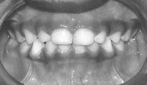 FIGURE 127.1. Normal nonspaced primary dentition with coincident midlines and overlap of maxillary teeth over the mandibular teeth. |
Normal Occlusion and Malocclusion
Occlusion refers to the manner in which the teeth fit together when biting and in the variety of tooth contacts that occur during mastication, swallowing, clenching, grinding, and other normal and abnormal mandibular movements. Occlusion is affected by the relative positions of the skeletal bases (the maxilla
and the mandible), by the position of the alveolar bone around the teeth, and by the relative positions of the teeth within the alveolar bone.
and the mandible), by the position of the alveolar bone around the teeth, and by the relative positions of the teeth within the alveolar bone.
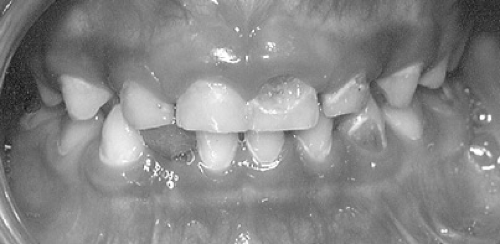 FIGURE 127.2. Primary dentition with white spot lesions, cavitated dental caries, and asymmetric, atypical loss of mandibular incisor. |
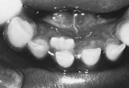 FIGURE 127.3. Double row of teeth in which a permanent incisor has erupted before primary tooth exfoliation. (Courtesy of Dr. Mark Wagner, University of Maryland Dental School.) |
In dentistry, every bite that differs from ideal occlusion is considered malocclusion. Malocclusion can be caused by skeletal or dental imbalance or by a combination of the two. Various degrees of malocclusion occur. Some malocclusion is considered within the normal range and is compatible with good dental health and function. Between 75% and 90% of children younger than 18 years in the United States have some degree of malocclusion. Approximately 15% to 30% have a handicapping condition requiring orthodontic treatment.
The significance of malocclusion is that it can interfere with chewing coarse or tough foods. It might not be aesthetically appealing, an important factor that causes psychological problems for some children. Malocclusion can cause trauma of soft tissues so severe that the tissue is stripped from the bone around teeth and the teeth are lost. Whereas some malocclusions can be corrected with early growth modification and some at a later age with orthodontics alone, some malocclusions are so severe that orthognathic surgery is required in addition to orthodontics.
The occlusal form of each dental arch should be smooth, symmetric, and without crowding or undesirable spacing. Lack of symmetry can indicate skeletal growth discrepancy, space loss due to trauma or caries, excessive rotation of teeth, or a congenitally missing tooth.
The dental midlines of the mandibular and maxillary arches should coincide with each other in occlusion (see Fig. 127.1) and should coincide with the midline of the face. Midline discrepancies greater than 1 mm indicate skeletal or dental problems that require dental referral.
Spacing in the primary dentition is generally desirable because the permanent teeth replacing the primary teeth are usually larger and require more space. Maxillary anterior spacing in the mixed dentition can be caused by pressure from the unerupted permanent canines on the incisor roots and may close on eruption of the canines. However, missing or impacted teeth or trauma can cause undesirable spaces in the mixed dentition. Spacing in the permanent dentition is generally not as desirable.
Spacing in the permanent dentition can be caused by generalized or localized smaller tooth structure than arch space, supernumerary teeth, a large fibrous frenum, congenitally missing (Fig. 127.4) or extracted teeth, cysts, and abnormal and unbalanced forces on the dentition such as those seen with thumb sucking. In some cases, adjacent teeth drift into the spaces through rotation and tipping, contributing to malocclusion.
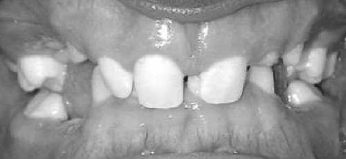 FIGURE 127.4. Multiple congenitally missing teeth and deep overbite in the mixed dentition of patient with ectodermal dysplasia. |
Crowding in the primary dentition is usually an indication that crowding will become worse with age. Slight crowding in the mixed dentition might be alleviated later.
The vertical relation between the permanent front teeth (i.e., overbite) is considered ideal when the maxillary incisors overlap the mandibular incisors about 1 to 2 mm (about 20% overlap). An overbite is considered severe if 5 to 7 mm of the mandibular incisors are covered; it is considered extreme if greater than 7 mm of the mandibular incisors are covered (Fig. 127.4). Approximately 15% to 20% of the children in the United States have severe to extreme overbites.
Teeth in the maxillary arch should hang outside those in the mandibular arch by approximately one-half of a tooth width. A crossbite exists if one or more maxillary teeth lie inside the mandibular teeth or completely outside the lower teeth (Fig. 127.5). Approximately 3% to 9% of the children in the United States have crossbites. Crossbites can be the result of dental positioning or skeletal discrepancies in the maxilla or the mandible. Small dental or skeletal discrepancies can cause the cusp tips of the teeth of both arches to meet and deflect to one side, resulting in a functional unilateral crossbite. Whereas small discrepancies are often difficult to detect, large discrepancies can result in noticeable facial asymmetry. Many patients with a unilateral crossbite exhibit facial asymmetry. Functional crossbites should be corrected as soon as possible to prevent increased asymmetry with age. Anterior crossbites in which the front maxillary teeth lie inside the mandibular teeth are frequently seen in children with concave or very straight faces, indicating skeletal imbalances.
A vertical open bite (Fig. 127.6) exists when the maxillary incisors do not touch the opposing incisors. Approximately 4% to 8% of adolescents in the United States have open bites. The
severity of the open bite can vary in the vertical height and in the number of teeth involved. Some children have an open bite that extends to their molars. For these children, chewing, such as biting through a sandwich, can be a problem. Open bites can be caused by digit sucking, excessive tooth mass for the jaw, trauma, and severe skeletal discrepancies. Children with skeletal open bites can have excessively long lower faces.
severity of the open bite can vary in the vertical height and in the number of teeth involved. Some children have an open bite that extends to their molars. For these children, chewing, such as biting through a sandwich, can be a problem. Open bites can be caused by digit sucking, excessive tooth mass for the jaw, trauma, and severe skeletal discrepancies. Children with skeletal open bites can have excessively long lower faces.
 FIGURE 127.5. Functional unilateral crossbite with noncoincident midlines in the primary dentition. |
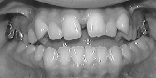 FIGURE 127.6. Vertical open bite and posterior crossbite in the permanent dentition. |
Normally, the backs of the maxillary incisors should touch the front of the mandibular incisors. If a horizontal open bite exists between the maxillary and mandibular incisors, it is called an overjet. An overjet can result from problems such as a discrepancy in the lengths of the maxilla and mandible or digit habits. A child with a large overjet can have an excessively convex profile because of a relatively deficient mandible as compared with the maxilla.
All posterior maxillary teeth should touch the mandibular teeth unless the patient is at a normal dental developmental stage in which primary teeth are exfoliating and permanent teeth are erupting. The lack of occlusion or the presence of vertical space may indicate a growth discrepancy in the area. Eruption proceeds at approximately 1 mm a month, and a space caused by a normal exfoliation and eruption of teeth should be closed or almost closed within approximately 6 months. Occasionally, teeth are partially visible but impacted because of lack of arch space.
In general, a child with crowding, a crossbite, an open bite, midline discrepancies, a deep bite, or a large overjet should be referred to an orthodontist for evaluation.
Determining the anteroposterior relation of the maxillary teeth to the mandibular teeth can be difficult. The key to screening a patient for gross occlusal problems is that the profile frequently reflects the relation of the maxilla to the mandible. A young child normally has a slightly convex facial profile that becomes straighter with growth of the mandible. Teenagers who have a maxilla and a mandible that are in good relation to each other tend to have straight profiles. A profile that is definitely convex or concave indicates skeletal imbalances, and orthodontic referral is recommended for those patients. Early intervention in the growth processes of the maxilla or mandible may prevent future surgical procedures to position the jaws better. However, mandibular growth can be difficult to predict. Mandibular growth can continue into adulthood, requiring surgery to correct the facial deformity and related malocclusion.
HABITS THAT CONTRIBUTE TO ORAL PROBLEMS
Habits such as digit sucking and lip sucking can contribute to malocclusions and, in the case of mouth breathing, to gingival inflammation. Other habits, such as bruxism and self-mutilation, cause destruction of oral tissues. However, some oral habits are little more than nuisances.
Digit Sucking
Digit sucking, which usually begins during the first year of life or before weaning, is the most common oral habit. Although the habit usually diminishes in frequency with age, some adults continue to suck their digits. A wide range exists in the reported prevalence, reaching a level as high as 86% of children between 1 and 10 years of age.
Possible causes of digit sucking include the rooting reflex, lack of sucking satisfaction during eating, peer modeling, and psychological problems. Although the habit is considered normal during infancy, the older child who continues to suck may have an emotional problem. Peer pressure and highly critical parents often compound the problem.
Digit sucking is a dental and social concern when it detrimentally affects occlusion. Many children discontinue the habit early or do not suck frequently or with great intensity. However, when the habit is frequent and intense, a greater chance exists that significant dental and skeletal deformities will be present. The deformities include anterior open bite, flaring maxillary incisors, retruded and crowded mandibular incisors, increased overjet, posterior crossbite, anteriorly displaced maxilla, and retruded mandible.
The critical age at which digit sucking should be stopped to minimize the effect on the permanent dentition is controversial. Many open bites self-correct if a child stops sucking before eruption of the maxillary permanent anterior teeth. Self-correction of the malocclusion depends on its severity, the flaccidity of the perioral soft tissue, and the presence of other oral habits, such as tongue position, mouth breathing, and lip habits. Severe oral problems require early intervention by the dentist.
Digit sucking can involve the thumb or one or more fingers. A variety of positions for the digits are assumed during sucking and appear to cause different occlusal changes. For example, a child who sucks only a digit on one side may exhibit a one-sided open bite. In contrast, a two-thumb sucker usually exhibits a wide and more symmetric open bite.
Even if the clinician does not see the child sucking and the parents do not report the habit, digit sucking should be considered if an open bite or overjet is observed during oral examination. If the incisors involved in the overjet are spaced and have no lingual support from the mandibular incisors, the clinician should suspect a digit-sucking habit. Children frequently do not respond or are untruthful to direct questions about sucking. Instead of asking directly, the hands of the child should be examined for extra clean, wrinkled, or red digits and calluses, which are diagnostic of frequent, intense sucking. The physician can then ask less threatening questions: “Are these the fingers that you suck the most?” “Do these fingers taste the best?” In the absence of signs on the hands, the examiner can ask, “Which fingers do you like to suck the most?” to elicit more truthful answers.
A simple explanation of the effects of the habit on the teeth may help some children stop their habit. Before deciding on any definitive treatment, however, the physician should determine the child’s desire to stop. If the child is motivated, positive reinforcement programs with the parents’ cooperation can be established. A reward system or a reminder such as an adhesive bandage on the digit can be used. If the habit is too deeply established to stop by positive reinforcement alone, the dentist can insert an intraoral habit appliance to serve as a reminder. This is usually effective. Additional orthodontics may be required in some children.
If the child is not motivated to stop the habit, appliances should not be used. The child may continue to suck, embedding the appliance into the soft tissue or causing orthopedic movement of the maxilla or intrusion of abutment teeth. Alternatively, the child may cause tissue damage by removing the fixed appliance. Counseling should be suggested to determine the reason for the child’s lack of motivation.
If digit sucking is associated with an emotional problem, counseling should be encouraged. Counseling should be considered for parents or families who cannot cope with the child’s habit. Negative reinforcement of the habit causes some children to become more adamant about sucking.
Pacifier Sucking
Prolonged and intense sucking of a pacifier can cause malocclusions similar to those produced by digit sucking. The problems are usually minimal and tend to self-correct after the habit is discontinued.
Pacifiers are used to satisfy an infant’s nonnutritive sucking needs and delay an infant’s feeding time when nursing or bottle-feeding is inconvenient. Prevalence studies report that as many as 45% of infants use pacifiers. The habit is discontinued in most children by 3 years of age, but it should be discontinued by 1 year. The simplest form of treatment is to discard the pacifier so that the child cannot find it. Parents should be cautioned not to dip pacifiers in honey or other sweet liquids, a practice associated with rampant caries.
Lip Habits
The two major lip habits involve wedging the lips between prominent upper incisors and the lower incisors and licking, sucking, or biting the lips. Forcefully wedging the lower lip between the teeth can cause additional protrusion of the upper incisors. Puckering of the skin over the chin occurs during this activity, because the mentalis muscle inserts into the soft tissue of the chin. An intraoral appliance can be used to minimize the action, but it does not correct the malocclusion.
Licking, sucking, or biting the lips is not associated with malocclusion but may result in chapping or drying of the lips and surrounding skin. Lip balm, face cream, or other lubricating material is recommended for palliative treatment.
Bruxism
Bruxism refers to the grinding of the teeth. The maxillary and mandibular teeth normally contact only during chewing and swallowing. During most of the day, they assume a rest position with as much as 5 mm of interocclusal space between the two arches. However, some children clench or grind their teeth.
The clinical signs vary from small wear facets to extensive wear of the teeth. The abrasion appears to stimulate cells within the pulp to form additional (sclerotic) dentin to protect the pulp. In some cases, the rate of abrasion is so great that the pulp can be seen through clear sclerotic dentin. In severe cases, the rate of abrasion exceeds the rate of dentin formation, exposing the pulp and resulting in a dental abscess. Bruxism can contribute to fracture of the teeth, muscle fatigue, and temporomandibular joint dysfunction and discomfort.
Bruxism is usually a subconscious activity and may occur during waking or sleeping periods. Parents usually report that the child grits his or her teeth together, particularly at night. Children with neurologic disorders are reported to engage in bruxism with the same intensity day and night.
If crossbites are observed, orthodontic treatment is indicated. If psychological stress is contributing to the bruxism, parental and child counseling and psychiatric referral may be necessary. Bite guards can provide palliative treatment when worn at night and, if necessary, during the day.
Mouth Breathing
Mouth breathing is associated with excessive drying of the anterior gingiva with a concomitant increase in chronic gingivitis. This effect is seen in patients who cannot close their lips easily or whose normal rest position of the lips is open.
The causal association between mouth breathing and a facial type characterized by a long, narrow face, short, flaccid lips, a narrow nose, and an expressionless face is controversial. It would be logical to expect factors such as adenoidal hypertrophy and allergy to affect a child with narrow nasal passages more than a child with wide passages, but some children with open-mouth posture and a mouth-breathing habit have no history of significant nasal obstruction. In addition, some children whose nasal obstruction is eliminated continue to mouth breathe.
Orthodontic treatment can eliminate the malocclusion and minimize some factors involved in not being able to close the lips properly. Evaluation by an otolaryngologist and an allergist may be necessary. If the mouth breathing continues despite a patent nasal airway, a program of positive reinforcement or the use of an oral shield over the lips may be effective.
Tongue Thrust
Tongue thrust is an infantile pattern of swallowing in which the tongue flattens and moves forward between the anterior teeth. Approximately 97% of newborns exhibit tongue thrust. Tongue thrust decreases with age; 3% of 12-year-old children exhibit the habit.
Tongue thrust is associated with open bite, incisor protrusion, and mandibular retrusion. However, the relation is uncertain. Three-fourths of children with tongue thrust who exhibit malocclusion in the primary teeth do not develop malocclusion of the permanent teeth. The swallowing pattern appears to mature in most children by 8 or 9 years of age, the time in which the permanent incisors are completely erupted. The efficacy of intraoral appliances is documented, but not always permanent. Treatment is contraindicated if no malocclusion or speech problem exists.
The cause of tongue thrust is controversial. Functionally, it appears that the tongue compensates for a small jaw and large lymphoid tissue by anterior thrust during swallowing. Growth of the mandible and reduction of lymphoid tissue appear to correspond with decreased thrust.
Intraoral and Perioral Piercings
Placing jewelry into a pierced tongue or lip is associated with tooth fractures, scarring, metal hypersensitivities, and infections. Piercings should be discouraged.
ORAL ANOMALIES
Various oral anomalies associated with developmental disturbances such as clefts can be detected at birth, within a few weeks of birth, or at the time of tooth eruption, whereas others such as ectodermal dysplasia are usually diagnosed much later. The categorization of oral anomalies into discrete entities is difficult since some are manifestations of an entire tissue
dysfunction, such as ectodermal dysplasia, whereas others are nonspecific. Disorders involving mineralization, such as vitamin D–resistant rickets, can affect bone and tooth formation. Although abnormalities of the teeth can be differentiated simplistically on the basis of tooth color, shape, number, position, and eruption, this method of categorization has its pitfalls, and frequently overlaps exist. Understanding of the genetic and environmental basis of numerous conditions (e.g., congenitally missing teeth) is increasing, but is often rudimentary.
dysfunction, such as ectodermal dysplasia, whereas others are nonspecific. Disorders involving mineralization, such as vitamin D–resistant rickets, can affect bone and tooth formation. Although abnormalities of the teeth can be differentiated simplistically on the basis of tooth color, shape, number, position, and eruption, this method of categorization has its pitfalls, and frequently overlaps exist. Understanding of the genetic and environmental basis of numerous conditions (e.g., congenitally missing teeth) is increasing, but is often rudimentary.
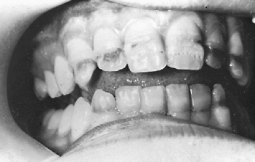 FIGURE 127.7. Linear hypoplasia resulting from tetracycline treatment at approximately 2 to 3 years of age. |
The pediatrician should know that different tooth types undergo formation at different times and that crown formation starts at different times for the permanent teeth compared with the primary teeth. The crowns of primary teeth and the first permanent molars begin formation in utero, whereas the other permanent teeth usually mineralize after birth. Knowledge of the effects of genetic disorders such as ectodermal dysplasia (see Fig. 127.4), drugs such as tetracycline (Fig. 127.7), and treatments such as head and neck irradiation (Fig. 127.8) on tooth formation allows the clinician to counsel parents about their children’s future dental development and to assess the risks and benefits of treatment. More than 200 syndromes are associated with dental or head and neck anomalies. However, only clefts will be discussed because of the lack of space.
Clefts
Incomplete or total lack of fusion of the various facial processes during the fifth to seventh week in utero can result in various forms of clefting. Clefting in the maxillary area is far more common than clefting of the lower lip or jaw and can involve different structures. Approximately 1 in every 600 newborn babies worldwide is affected by cleft lip with or without cleft palate (CL/P) or isolated cleft palate (CP). The prevalence of CL/P reportedly varies depending on the ethnic group and the manner of reporting.
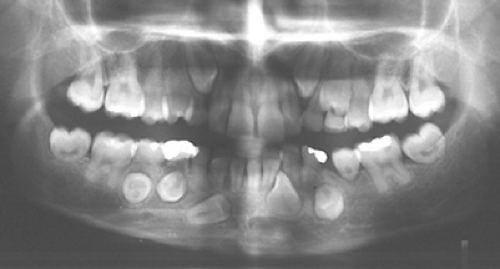 FIGURE 127.8. Panoramic radiograph showing lack of permanent mandibular tooth root formation and tooth impaction caused by two bouts of radiation treatment before the age of 5 years. |
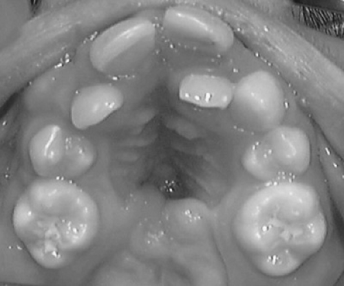 FIGURE 127.9. Maxillary arch of patient with a partially repaired isolated cleft palate showing malocclusion with severe crowding, congenitally missing teeth, and malalignment. |
The severity of the cleft and its associated oral problems varies from an incomplete cleft lip with only a small notching in the vermillion border to a complete cleft lip, either unilateral or bilateral, that extends through the alveolar process affecting the dentition and the dental arch form. Isolated CP can involve only the uvula or extend through the hard palate (Fig. 127.9).
Clefting can be manifested as part of a syndrome caused by single mutant genes or by chromosomal defects such as trisomy 13. Environmental factors such as maternal anticonvulsant drugs and smoking are implicated in some cases.
The patient with clefts may have numerous problems in addition to the cosmetic appearance. Palatal clefting may affect an infant’s ability to feed because of interference with sucking. Palatal clefting also affects the child’s speech.
Abnormalities in tooth number, structure, and appearance frequently occur in the area of clefting. Missing teeth, supernumerary teeth, and malformed teeth are common in the areas surrounding the cleft. Supernumerary and malformed teeth will need to be evaluated for extraction or restoration. Radiographs are necessary to determine the status of these teeth prior to alveolar grafting. The alveolar bone in the area of the cleft is often inadequate to support adjacent erupting teeth.
Stay updated, free articles. Join our Telegram channel

Full access? Get Clinical Tree


