Risk Factors and Prevention
The principal risk is the large baby, but only about half of all cases occur in babies over 4 kg. Further, antenatal prediction of fetal size, even with ultrasound, is poor. Other reported factors include previous shoulder dystocia, increased maternal body mass index (BMI), labour induction, low height, maternal diabetes and instrumental delivery. Antenatal prediction (AmJOG 2006; 195: 1544) is limited by the poor sensitivity even of integration [→ p.153] of these risk factors, coupled with the rarity of a serious outcome and the fact that prevention involves Caesarean section. Most cases are therefore considered unpreventable.
Management
This requires rapid and skilled intervention: teaching of this rather than attempted prevention is current practice. A sequence of actions is recommended. Because the obstruction is at the pelvic inlet, excessive traction is useless, and will cause Erb’s palsy: gentle downward traction is used. Initially, senior help is requested, and the legs are hyperextended onto the abdomen (McRoberts’ manoeuvre); suprapubic pressure is also applied. These methods work in about 90% of cases. If they fail, internal manoeuvres are required, necessitating episiotomy. If the shoulders are transverse, pressure behind the anterior shoulder will rotate it to the widest diameter; this can be combined with pressure on the anterior part of the posterior shoulder (Wood’s screw manoeuvre). If this fails, the posterior arm is grasped and, by extension at the elbow, the hand is brought down. The trunk will either follow, or rotation of the body using the arm is performed, causing the anterior shoulder to enter the pelvis. Last resorts include symphisiotomy, after lateral replacement of the urethra with a metal catheter, and the Zavanelli manoeuvre. This involves replacement of the head and Caesarean section, but by this time fetal damage is usually irreversible.
Cord Prolapse
Definition and Consequences
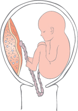
This occurs when, after the membranes have ruptured, the umbilical cord descends below the presenting part (Fig. 32.2). Untreated, the cord will be compressed or go into spasm and the baby will rapidly become hypoxic. It occurs in 1 in 500 deliveries.
Risk Factors and Prevention
Risks include preterm labour, breech presentation, polyhydramnios, abnormal lie and twin pregnancy. More than half occur at artificial amniotomy. The diagnosis is usually made when the fetal heart rate becomes abnormal and the cord is palpated vaginally, or if it appears at the introitus. The widespread practice of delivering breeches by Caesarean section has reduced the incidence.
Management
Initially, the presenting part must be prevented from compressing the cord: it is pushed up by the examining finger, or tococlytics such as terbutaline are given. If the cord is out of the introitus, it should be kept warm and moist but not forced back inside. The patient is asked to go on ‘all fours’, whilst preparations for delivery by the safest route are undertaken. Immediate Caesarean section is normally used, but instrumental vaginal delivery is appropriate if the cervix is fully dilated and the head is low. With prompt treatment, fetal mortality is rare.
Amniotic Fluid Embolism
Definition and Consequences
Stay updated, free articles. Join our Telegram channel

Full access? Get Clinical Tree


