Chapter 584 Neurologic Evaluation
A comprehensive neurologic evaluation—including history, physical examination, and the judicious use of ancillary studies—allows the clinician to localize and determine the etiology of central and peripheral nervous system pathology.
History
A detailed history is the cornerstone of any neurologic assessment. Although parents may be the primary informants, most children older than 3-4 yr are capable of contributing to their history and should be questioned directly.
The history should begin with the chief complaint, as well as a determination of the complaint’s relative significance within the context of normal development (Chapters 7–14). The latter step is critical because a 13 mo old who cannot walk may be perfectly normal, whereas a 4 yr old who cannot walk might have a serious pathology.
Following the chief complaint and history of present illness, the physician should obtain a complete birth history, particularly if a congenital disorder is suspected. The birth history should begin with a review of the pregnancy, including specific questions about common complications, such as pregnancy-induced hypertension, preeclampsia, gestational diabetes, vaginal bleeding, infections, and falls. It is important to quantify any cigarette, alcohol, or drug (prescription, herbal, illicit) use. Inquiring about fetal movement might provide clues to an underlying diagnosis, because decreased or absent fetal activity can be associated with chromosomal anomalies and CNS or neuromuscular disorders. Finally, any abnormal ultrasound or amniocentesis results should be noted.
A labor history should address the gestational age at delivery and mode of delivery (spontaneous vaginal, vacuum- or forceps-assisted, cesarean section) and should comment on the presence or absence of fetal distress. If delivery was by cesarean section, it is essential to record the indication for surgery.
The birth weight, length, and head circumference provide useful information about the duration of a given problem, as well as insights into the uterine environment. Parents can usually provide a reliable history of their child’s postnatal course; however, if the patient was resuscitated or had a complicated hospital stay, it is often helpful to obtain the hospital records. The physician should inquire about the infant’s general well-being, feeding and sleeping patterns, activity level, and the nature of his or her cry. If the infant had jaundice, it is important to determine both the degree of jaundice and how it was managed. Historical markers of neurologic dysfunction include full-term infants who are unable to breathe spontaneously; have poor, uncoordinated sucks; need an inordinate amount of time to feed; or require gavage feeding. Again, it is important to consider the developmental context, because all of these issues would be expected in premature infants, particularly those with very low birth weights.
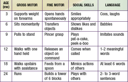
The most important component of a neurologic history is the developmental assessment (Chapters 6 and 14). Careful evaluation of a child’s social, cognitive, language, fine motor, and gross motor skills is required to distinguish normal development from either isolated or global (i.e., in two or more domains) developmental delay. An abnormality in development from birth suggests an intrauterine or perinatal cause, but a loss of skills (regression) over time strongly suggests an underlying degenerative disease of the CNS, such as an inborn error of metabolism. The ability of parents to recall the precise timing of their child’s developmental milestones is extremely variable. It is often helpful to request old photographs of the child or to review the baby book, where the milestones may have been dutifully recorded. In general, parents are aware when their child has a developmental problem, and the physician should show appropriate concern. Table 584-1 outlines the upper limits of normal for attaining specific developmental milestones. A comprehensive review of developmental screening tests and their interpretation is listed in Chapter 14.
Family history is extremely important in the neurologic evaluation of a child. Most parents are extremely cooperative in securing medical information about family members, particularly if it might have relevance for their child. The history should document the age and history of neurologic disease, including developmental delay, epilepsy, migraine, stroke, and inherited disorders, for all first- and second-degree relatives. It is important to inquire directly about miscarriages or fetal deaths in utero and to document the sex of the embryo or fetus, as well as the gestational age at the time of demise. When available, the results of postmortem examinations should be obtained, as they can have a direct bearing on the patient’s condition. The parents should be questioned about their ethnic backgrounds, because some genetic disorders occur more commonly within specific populations (e.g., Tay-Sachs in the Ashkenazi Jewish population). They should also be asked if there is any chance that they could be related to each other, because the incidence of metabolic and degenerative disorders of the CNS is increased significantly in children of consanguineous marriages.
The social history should detail the child’s current living environment, as well as his or her relationship with other family members. It is important to inquire about recent stressors, such as divorce, remarriage, birth of a sibling, or death of a loved one, because they can affect the child’s behavior. If the child is in daycare or school, one should document his or her academic and social performance, paying particular attention to any abrupt changes. Academic performance can be assessed by asking about the child’s latest report card, and peer relationships can be evaluated by having the child name his or her “best friends.” Any child who is unable to name at least 2 or 3 playmates might have abnormal social development. In some cases, discussions with the daycare worker or teacher provide useful ancillary data.
Neurologic Examination
The neurologic examination begins at the outset of the interview. Indirect observation of the child’s appearance and movements can yield valuable information about the presence of an underlying disorder (Chapters 6 and 14). For instance, it may be obvious that the child has dysmorphic facies, an unusual posture, or an abnormality of motor function manifested by a hemiparesis or gait disturbance. The child’s behavior while playing and interacting with his or her parents may also be telling. A normal child usually plays independently early in the visit but will rapidly engage in the interview process. A child with attention-deficit/hyperactivity disorder might display impulsive behavior in the examining room, and a child with neurologic impairment might exhibit complete lack of awareness of the environment. Finally, note should be made of any unusual odors about the patient, because some metabolic disorders produce characteristic scents (e.g., the “musty” smell of phenylketonuria or the “sweaty feet” smell of isovaleric acidemia). If such an odor is present, it is important to determine whether it is persistent or transient, occurring only with illnesses.
The examination should be conducted in a nonthreatening, child-friendly setting. The child should be allowed to sit where he or she is most comfortable, whether it be on a parent’s lap or on the floor of the examination room. The physician should approach the child slowly, reserving any invasive or painful tests (e.g., measurement of head circumference, gag reflex) for the end of the examination. In the end, the more that the examination seems like a game, the better the child will cooperate. Because the neurologic examination of an infant requires a somewhat modified approach from that of an older child, the two groups are considered separately (Chapters 7, 8, and 88).
Mental Status
Age aside, the neurologic examination should include an assessment of the patient’s mental status in terms of both level of arousal and interaction with the environment. Premature infants born at <28 wk of gestation do not have consistent periods of alertness, whereas slightly older infants arouse from sleep with gentle physical stimulation. Sleep-wake patterns are well developed at term. Because the level of alertness of a neonate depends on many factors, including the time of the last feeding, room temperature, and gestational age, serial examinations are critical when evaluating for changes in neurologic function. Older children’s mental status can be assessed by watching them play. Having them tell a story, draw a picture, or complete a puzzle can also be helpful in assessing cognitive function. Memory can be evaluated informally as patients recount their personal information, as well as more formally by asking them to register and recall three objects or perform a digit span.
Head
Correct measurement of the head circumference is important. It should be performed at every visit for patients younger than 3 yr and should be recorded on a suitable head growth chart. To measure, a nondistensible plastic measuring tape is placed over the mid-forehead and extended circumferentially to include the most prominent portion of the occiput. If the patient’s head circumference is abnormal, it is important to document the head circumferences of the parents and siblings. Errors in the measurement of a newborn skull are common owing to scalp edema, overriding sutures, and the presence of cephalohematomas. The average rate of head growth in a healthy premature infant is 0.5 cm in the 1st 2 wk, 0.75 cm in the 3rd wk, and 1.0 cm in the 4th wk and every week thereafter until the 40th wk of development. The head circumference of an average term infant measures 34-35 cm at birth, 44 cm at 6 mo, and 47 cm at 1 yr of age (Chapters 7 and 8).
If the brain is not growing, the skull will not grow; therefore, a small head reflects a small brain, or microcephaly. Conversely, a large head may be associated with a large brain, or macrocephaly, which is most commonly familial but may be due to a disturbance of growth, neurocutaneous disorder (e.g., neurofibromatosis), chromosomal defect (e.g., Kleinfelter syndrome), or storage disorder. Alternatively, the head size may be increased secondary to hydrocephalus (Fig. 584-1) or chronic subdural hemorrhages. In the latter case, the skull tends to assume a square or boxlike shape, because the long-standing presence of fluid in the subdural space causes enlargement of the middle fossa.
The shape of the head should be documented carefully. Plagiocephaly, or flattening of the skull, can be seen in normal infants but may be particularly prominent in hypotonic or weak infants, who are less mobile. A variety of abnormal head shapes can be seen when cranial sutures fuse prematurely, as in the various forms of inherited craniosynostosis (Chapter 585.12).
Inspection of the head should include observation of the venous pattern, because increased ICP and thrombosis of the superior sagittal sinus can produce marked venous distention. Dysmorphic facial features can indicate a neurodevelopmental aberration. Likewise, cutaneous abnormalities, such as cutis aplasia or abnormal hair whorls, can suggest an underlying brain malformation or genetic disorder.
Palpation of a newborn’s skull characteristically reveals molding of the skull accompanied by overriding sutures—a result of the pressures exerted on the skull during its descent through the pelvis. Marked overriding of the sutures beyond the early neonatal period is cause for alarm, because it suggests an underlying brain abnormality. Palpation additionally might reveal bony bridges between sutures (craniosynostosis), cranial defects, or, in premature infants, softening of the parietal bones (craniotabes).
Auscultation of the skull is an important adjunct to the neurologic examination. Cranial bruits may be noted over the anterior fontanel, temporal region, or orbits and are best heard using the diaphragm of the stethoscope. Soft symmetric bruits may be discovered in normal children <4 yr of age or in association with a febrile illness. Demonstration of a loud or localized bruit is usually significant and warrants further investigation, because they may be associated with severe anemia, increased ICP, or arteriovenous malformations of the middle cerebral artery or vein of Galen. It is important to exclude murmurs arising from the heart or great vessels, because they may be transmitted to the cranium.
Cranial Nerves
Olfactory Nerve (Cranial Nerve I)
Anosmia, loss of smell, most commonly occurs as a transient abnormality in association with an upper respiratory tract infection or allergies. Permanent causes of anosmia include head trauma with damage to the ethmoid bone or shearing of the olfactory nerve fibers as they cross the cribiform plate, tumors of the frontal lobe, intranasal drug use, and exposure to toxins (acrylates, methacrylates, cadmium). Occasionally, a child who recovers from purulent meningitis or develops hydrocephalus has a diminished sense of smell. Rarely, anosmia is congenital, in which case it can occur as an isolated deficit or as part of Kallman syndrome, a familial disorder characterized by hypogonadotropic hypogonadism and congenital anosmia. Although not a routine component of the examination, smell can be tested reliably as early as the 32nd wk of gestation by presenting a stimulus and observing for an alerting response, withdrawal, or both. Care should be taken to use appropriate stimuli, such as coffee or peppermint, as opposed to strongly aromatic substances (e.g., ammonia inhalants) that stimulate the trigeminal nerve. Each nostril should be tested individually by pinching shut the opposite side.
Optic Nerve (Cranial Nerve II)
Assessment of the optic disc and retina is a critical component of the neurologic examination. Although the retina is best visualized by dilating the pupil, most physicians do not have ready access to mydriatic agents at the bedside; therefore, it may be necessary to consult an ophthalmologist in some cases. Mydriatics should not be administered to patients whose pupillary responses are being followed as a marker for impending herniation or to patients with cataracts. When mydriatics are used, both eyes should be dilated, because unilateral papillary fixation and dilation can cause confusion and worry in later examiners unaware of the pharmacologic intervention. Examination of an infant’s retina may be facilitated by providing a nipple or soother and by turning the head to one side. The physician gently strokes the patient to maintain arousal, while examining the closer eye. An older child should be placed in the parent’s lap and should be distracted by bright objects or toys. The color of the optic nerve is salmon-pink in a child but may be gray-white in a newborn, particularly if he or she has fair coloring. This normal finding can cause confusion and can lead to the improper diagnosis of optic atrophy.
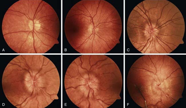
Disc edema refers to swelling of the optic disc, and papilledema specifically refers to swelling that is secondary to increased intracranial pressure. Papilledema rarely occurs in infancy because the skull sutures can separate to accommodate the expanding brain. In older children, papilledema may be graded according to the Frisen scale (Fig. 584-2). Disc edema must be differentiated from papillitis, or inflammation of the optic nerve. Both conditions manifest with enlargement of the blind spot, but visual acuity and color vision tend to be spared in early papilledema in contrast to what occurs in optic neuritis.

Figure 584-2 Stages of papilledema (Frisen scale). A, Stage 0: Normal optic disc. B, Stage 1: Very early papilledema with obscuration of the nasal border of the disc only, without elevation of the disc borders. C, Stage 2: Early papilledema showing obscuration of all borders, elevation of the nasal border, and a complete peripapillary halo. D, Stage 3: Moderate papilledema with elevation of all borders, increased diameter of the optic nerve head, obscuration of vessels at the disc margin, and a peripapillary halo with finger-like extensions. E, Stage 4: Marked papilledema characterized by elevation of the entire nerve head and total obscuration a segment of a major blood vessel on the disc. F, Stage 5: Severe papilledema with obscuration of all vessels and obliteration of the optic cup. Note also the nerve fiber layer hemorrhages and macular exudate.
(Photographs courtesy of the University of Rochester Eye Institute.)
Retinal hemorrhages occur in 30-40% of all full-term newborn infants. The hemorrhages are more common after vaginal delivery than after cesarean section and are not associated with birth injury or with neurologic complications. They disappear spontaneously by 1-2 wk of age. The presence of retinal hemorrhages beyond the early neonatal period should raise a concern for nonaccidental trauma.
Vision
At 28 wk of corrected gestational age, a premature infant blinks in response to a bright light, and at 32 wk, he or she maintains eye closure until the light source is removed. A normal 37-wk infant turns the head and eyes toward a soft light, and a term infant is able to fix on and follow a target, such as the examiner’s face. Optokinetic nystagmus (OKN), which is conjugate nystagmus that occurs during attempted fixation on a series of rapidly moving objects, can also be used as a crude assessment of the visual system in infants. OKN is elicited by moving an OKN tape—usually a strip of material with alternating 2-inch black and white strips—across the patient’s visual field. Although OKN responses can be tested monocularly in neonates, they do not become symmetric until 4-6 mo of age.
Visual fields can be tested in an infant or young child by advancing a brightly colored object from behind the patient’s head into the peripheral visual field and noting when he or she first looks at the object. Suspension of the object by a string prevents the patient from focusing on the examiner’s hand and arm. The examiner should be certain that the patient is responding to seeing, not hearing, the object.
Visual acuity in term infants approximates 20/150 and reaches the adult level of 20/20 by about 6 mo of age. Children who are too young to read the standard letters on a Snellen eye chart may learn the “E game,” which entails pointing to indicate the direction that the E is facing. Children as young as to 3 yr of age can identify the objects on a pediatric eye chart (Allen chart) at a distance of 15-20 ft.
The pupil reacts to light by 29-32 wk of corrected gestational age; however, the pupillary response is often difficult to evaluate, because premature infants resist eye closure and have poorly pigmented irises. Pupillary size, symmetry, and reactivity may be affected by drugs, space-occupying brain lesions, metabolic disorders, and abnormalities of the optic nerves and midbrain. A small pupil may be seen as part of the Horner syndrome—characterized by ipsilateral ptosis (droopy eyelid), miosis (constricted pupil), and anhidrosis (lack of sweating) of the face. Horner syndrome may be congenital or may be caused by a lesion of the sympathetic pathway in the hypothalamus, brainstem, cervical spinal cord, or sympathetic plexus. Localization of the lesion within the sympathetic nervous system may be obvious given the other signs present or may be uncertain. In the latter case, serial testing with cocaine drops followed by hydroxyamphetamine drops may be helpful.
During the examination of the pupil, any abnormalities of the iris should also be noted (e.g., heterochromia, Brushfield spots). The physician should also assess the posterior segment of the eye using the red reflex test, which is performed in a darkened room using a direct ophthalmoscope held close to the examiner’s eye and 12 to 18 inches from the infant’s eyes. If the posterior segment of the eye is normal, the examiner should see symmetric reddish-pink retinal reflections. The absence of any red reflex or the presence of a blunted reflex, white reflex (leukocoria), or red reflex with dark spots all signal pathology and should prompt referral to an ophthalmologist.
Oculomotor (III), Trochlear (IV), and Abducens Nerves (VI)
The globe is moved by 6 extraocular muscles, which are innervated by the oculomotor, trochlear, and abducens nerves. These muscles and nerves can be assessed by having the patient follow an interesting toy or the examiner’s finger in the 6 cardinal directions of gaze. The physician observes the range and nature (conjugate vs. dysconjugate, smooth vs. choppy or saccadic) of the eye movements, particularly noting the presence and direction of any abnormal eye movements. Premature infants >25 wk of gestational age and comatose patients can be evaluated using the oculocephalic (doll’s eye) maneuver, in which the patient’s head is quickly rotated to evoke reflex eye movements. If the brainstem is intact, rotating the patient’s head to the right causes the eyes to move to the left and vice versa. Similarly, rapid flexion and extension of the head elicits vertical eye movement.
Disconjugate gaze can result from extraocular muscle weakness; cranial nerve (CN) III, IV, or VI palsies; or brainstem lesions that disrupt the medial longitudinal fasciculus. Infants who are <2 mo old can have slightly disconjugate gaze at rest, with one eye horizontally displaced from the other by 1 or 2 mm (strabismus). Vertical displacement of the eyes, known as skew deviation, is always abnormal and requires investigation. Strabismus is discussed further in Chapter 615.
When there is a subtle eye movement abnormality, the red glass test may be helpful in localizing the lesion. To perform this test, a red glass is placed over one of the patient’s eyes and he or she is instructed to follow a white light in all directions of gaze. The child sees one red/white light in the direction of normal muscle function but notes a separation of the red and white images that is greatest in the plane of action of the affected muscle.
In addition to gaze palsies, the examiner might encounter a variety of adventitious movements. Nystagmus

Stay updated, free articles. Join our Telegram channel

Full access? Get Clinical Tree

