Fig. 1
Peripheral nerves of the upper extremity. A summary view of the brachial plexus organization and the paths of the ulnar, radial, and median nerves. The site of each plane transition, identified as where the nerves pierce through or traverse above or below a given anatomic structure, along the respective nerve’s course is represented (Reprinted from Neurology board review: an illustrated study guide. M Mowzoon, 2007, Rochester, MN: Mayo Clinic Scientific Press. Copyright by Taylor and Francis Group LLC Books. Reprinted with permission. License Id: 3254140720553)
The long thoracic nerve (C5, 6, 7), dorsal scapular nerve (C5), and innervation for the scalene muscles (C5, 6) originate from the nerve roots in the posterior cervical triangle. The long thoracic nerve innervates the serratus anterior, which is responsible for stabilizing the scapula to allow anteversion of the arm. The dorsal scapular nerve pierces the middle scalene and innervates the rhomboid muscles, which stabilize and adduct the scapula. The superior trunk gives rise the subclavius nerve (C5, 6) and the suprascapular nerve (C5, 6). The suprascapular nerve travels with the suprascapular artery and vein before innervating the supraspinatus and infraspinatus muscles which assist with arm abduction and external rotation. The subclavius nerve often travels to the subclavius muscle with the phrenic nerve (Tubbs et al. 2010).
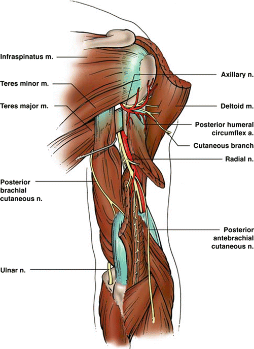
Emerging from the posterior cord are the upper subscapular (C5, 6), thoracodorsal (C5, 6, 7), lower subscapular (C5, 6), axillary (C5, 6), and radial nerves (C5, 6, 7, 8, T1). The thoracodorsal nerve follows the subscapular artery before innervating the latissimus dorsi muscle which produces arm adduction. The axillary nerve passes through the quadrangular space before dividing into an anterior and posterior branch with the anterior branch intimately winding around the surgical neck of the humerus (Fig. 2). The axillary nerve is mostly a motor nerve providing innervation to the deltoid, teres minor, and long head of triceps brachii muscles and therefore influences arm abduction, flexion, extension, and rotation (Tubbs et al. 2010). From the medial cord originate the medial pectoral nerve (C8, T1), medial brachial cutaneous nerve (C8, T1), medial antebrachial cutaneous nerve (C8, T1), a portion of the median nerve (C8, T1) and ulnar nerve (C8, T1). The medial pectoral nerve assists with arm adduction by always innervating the pectoralis minor and frequently the pectoralis major muscles. The medial brachial cutaneous nerve courses along the medial side of the proximal brachial artery and innervates the medial distal third of the arm. The medial antebrachial cutaneous nerve (MAC) also initially runs medial to the brachial artery but transitions to a superficial position in the middle arm as it runs with the basilic vein. In the antecubital fossa the MAC bifurcates to continue its venous relationship with the volar branch anterior to the median basilic vein and the ulnar branch posterior to the median vein. The MAC innervates the volar distal third of the arm and the ulnar half of both volar and dorsal surfaces of the forearm (Tubbs et al. 2010).


Fig. 2
Posterior view of the arm. The axillary nerve passes through the quadrangular space and then wraps around the surgical neck of the humerus with the posterior humeral circumflex artery. Note the early strong relationship between the radial nerve and the brachial artery as they enter the arm and travel along the spiral groove of the humerus. The medial and lateral heads of the triceps are resected but can be seen receiving radial innervation (Reprinted with permission from “Anatomy and landmarks for branches of the brachial plexus: a vade mecum,” by S.R. Tubbs et al. 2010, Surgical and Radiologic Anatomy, 32(3), p. 265. Copyright 2010 by Springer-Verlag France. Artist David License Id: 3244201311409)
The lateral pectoral nerve (C5, 6, 7) and musculocutaneous nerve (C5, 6, 7), and the remaining contribution to the median nerve (C5, 6, 7) arise from the lateral cord (Jacobson et al. 2010; Tubbs et al. 2010). The lateral pectoral nerve innervates the pectoralis major muscle also contributing to arm adduction. The musculocutaneous nerve penetrates and innervates the coracobrachialis muscle and then traverses the remainder of the arm radially between the biceps brachii and brachialis muscles, both of which receive innervation. After the motor innervation is exhausted, the nerve becomes the lateral antebrachial cutaneous nerve which runs under the bicipital aponeurosis before bifurcating and innervates the radial half of the volar forearm. The anterior branch of the lateral antebrachial cutaneous nerve terminates at the base of the thumb after traversing anteriorly over the radial artery at the wrist (Tubbs et al. 2010).
The ulnar nerve proceeds down the arm parallel and posterior to the brachial artery close to the humerus until the distal third of the arm where it then turns dorsally away from the artery. The nerve penetrates the medial intermuscular septum and lies against the medial head of the triceps muscle (Jacobson et al. 2010). The ulnar nerve will then pass under the arcade of Struthers, a fibrous band originating from the medial head of the triceps and inserting onto the medial intermuscular septum. The arcade of Struthers is present in approximately 70 % of the population but more likely to be altogether absent in younger pediatric patients (Feinberg et al. 1997). At the elbow the ulnar nerve lies posterior to the medial epicondyle of the humerus and continues under the cubital tunnel retinaculum, a short, thin fibrous band traversing between the medial epicondyle and the olecranon (Fig. 1). The cubital tunnel is immediately distal to the retinaculum with the floor formed by the ulna and the roof by Osborne’s ligament which bridges the two heads of the flexor carpi ulnaris (FCU) (Jacobson et al. 2010). The anconeus epitrochlearis, an established variant present in up to one third of the population, is an accessory muscle proximal to the cubital tunnel that inserts between the medial epicondyle and the olecranon and overlies the ulnar nerve parallel to Osborne’s fascia (Jacobson et al. 2010). In addition to the cubital tunnel itself, the anconeus epitrochlearis may be a source of ulnar nerve compression (Stutz et al. 2012). After exiting the cubital tunnel, the ulnar nerve lies between the FCU and flexor digitorum profundus (FDP) muscle bellies (Figs. 1 and 3). It is here that the nerve innervates the FCU and the ulnar muscle bellies of the FDP controlling the ring and little fingers (Jacobson et al. 2010). At or just proximal to the level of the wrist, two sensory nerves originate: the ulnar palmar cutaneous nerve providing sensation for the hypothenar eminence and the dorsal cutaneous nerve which provides sensation to the ulnar dorsal surface of the hand, the dorsal little finger, and a variable portion of the dorsal ring finger (Engber and Gmeiner 1980; Tagliafico et al. 2012).


Fig. 3
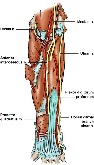
Anterior view of the forearm. In the distal arm, the median nerve is ulnar to the brachial artery, and after traversing the elbow, the anterior interosseous nerve is closely associated with the respective artery. In the mid-forearm the ulnar nerve becomes closely associated with the ulnar artery as it travels ulnarly between the flexor digitorum profundus and the flexor carpi ulnaris (cut) (Reprinted with permission from “Anatomy and landmarks for branches of the brachial plexus: a vade mecum,” by S.R. Tubbs et al. 2010, Surgical and Radiologic Anatomy, 32(3), p. 265. Copyright 2010 by Springer-Verlag France. Artist David Fisher. License Id: 3244201311409)
Distally the ulnar nerve joins the ulnar artery and enters Guyon’s canal, a tunnel formed by the pisiform bone, flexor retinaculum, and the palmar carpal ligament. Variably within the canal or shortly after exiting, the nerve either bifurcates or trifurcates into primarily sensory or motor nerves. The deeper motor branch passes under the flexor digiti minimi brevis and abductor digiti minimi to supply the hypothenar muscles, the deep head of flexor pollicis brevis, the adductor pollicis, the dorsal and palmar interossel, and the third and fourth lumbrical muscles (Jacobson et al. 2010; Tagliafico et al. 2012; Tubbs et al. 2010). The superficial sensory portion innervates the palmar surface of the distal ulnar palm and form the proper digital nerves to the little finger and ulnar half of the ring finger (Fig. 1) (Tagliafico et al. 2012).
In the arm the radial nerve travels diagonally in an ulnar to radial direction along the posterior aspect of the humerus in the spiral groove (Jacobson et al. 2010). Proximally the nerve innervates the medial and lateral heads of the triceps brachii muscle and the anconeus muscle, in addition to emitting several sensory nerves to provide sensation to the posterior arm and forearm: the posterior cutaneous nerve, the lower lateral cutaneous nerve, and the posterior cutaneous nerve of the forearm (Figs. 1 and 2). In the distal arm, the radial nerve penetrates the lateral intermuscular septum and courses under the brachioradialis muscle (Jacobson et al. 2010; Tubbs et al. 2010). Here the brachioradialis and extensor carpi radialis brevis and longus muscles receive their innervation (Tagliafico et al. 2012). After passing the elbow joint level at the lateral epicondyle, the radial nerve enters the anterior compartment of the forearm between the brachialis, brachioradialis, and extensor carpi radialis longus muscles before bifurcating into the superficial radial nerve and the deeper posterior interosseous nerve (PIN).
The PIN travels posteriorly through the arcade of Frohse between the two heads of the supinator and then courses in an intramuscular path close to the radius (Jacobson et al. 2010) (Fig. 1). The PIN is responsible for providing the innervation to the entire extensor compartment including the brachioradialis, supinator, extensor digitorum communis, extensor carpi ulnaris, extensor digiti minimi, abductor pollicis longus, abductor pollicis brevis, extensor pollicis longus (EPL), and extensor indicis muscles. While the PIN does not provide any cutaneous innervation, it does provide sensory feedback from the dorsal wrist capsule, through the terminal sensory branch, which travels posterior to the posterior interosseous membrane and anterior to the EPL (Smith et al. 2011; Tubbs et al. 2010).
The superficial branch of the radial nerve briefly follows the radial artery before coursing laterally in proximity to the cephalic vein and then through the anatomic snuffbox. The superficial branch is entirely sensory and innervates the radial aspect of the dorsum of the hand, the dorsum of thumb, index, long, and radial half of the ring fingers proximal to the distal interphalangeal joint (Jacobson et al. 2010; Tagliafico et al. 2012).
The median nerve descends down the arm adjacent to the brachial artery, generally lateral to the artery proximally, and then becomes medial to the artery distally (Fig. 3) (Jacobson et al. 2010). The median nerve traverses the elbow joint level adjacent to the origin of the pronator teres humeral head and then typically goes under this muscle belly, although a less common course is intramuscular through the pronator teres or brachialis muscles (Jacobson et al. 2010). Along the forearm the median nerve courses between the FDP and flexor digitorum superficialis (FDS) (Klauser et al. 2010). The anterior interosseous nerve (AIN) branches off radially after the median nerve crosses the two heads of the pronator teres and enters the FDP muscle belly before continuing on the volar surface of the interosseous membrane in proximity to the interosseous artery (Chin and Meals 2004). The AIN innervates the flexor pollicis longus, FDP of the index and middle fingers, and quadratus muscles. The AIN terminates as sensory innervation for the radiocarpal, midcarpal, and carpometacarpal joints (Chin and Meals 2004). The median nerve proper will innervate the pronator teres, flexor carpi radialis, FDS, and palmaris longus. Alternatively, the FDS may be innervated by the AIN, a reported variant, manifesting in weakness of all proximal interphalangeal joints following an isolated AIN injury (Chin and Meals 2004). Proximal to the carpal tunnel, the median nerve gives off the median palmar cutaneous branch which innervates the thenar eminence and the palmar triangle. At the wrist, the median nerve enters the carpal tunnel, which is formed by the carpus on the floor and the transverse carpal ligament as the roof (Figs. 1 and 3). Within the tunnel the median nerve lies superficial and radial, surrounded by the FPL radially and both FDP and FDS tendons of the index finger dorsally and ulnarly (Klauser et al. 2010). After exiting the carpal tunnel, the median nerve bifurcates into a smaller radial branch and a larger ulnar branch. The radial branch first gives off the recurrent motor branch for the thenar muscles and then trifurcates into three proper palmar digital nerves, two to the thumb and one to the radial side of the index finger. The recurrent motor nerve innervates the opponens pollicis, abductor pollicis brevis, and the superficial head of the flexor pollicis brevis muscles. The proper palmar digital nerves provide innervation to the entire palmar surface of the thumb and radial aspect of the index finger – with the index finger nerve also innervating the first lumbrical. The larger ulnar branch of the median nerve further bifurcates into two common palmar digital nerves, with the first common nerve innervating the second lumbrical and forming the proper digital nerves for the ulnar index finger and radial middle finger. The other common palmar digital nerve innervates the ulnar middle and radial ring fingers after dividing into the respective proper digital nerves. A dorsal branch from each proper digital nerve communicates with the digital nerves of the superficial radial branches to innervate the dorsal distal phalanx of the index, middle, and radial half of the ring fingers (Tagliafico et al. 2012).
It is important to remember that the aforementioned sensory and motor assignments are typical but not the rule as variations are commonplace. One of the more common variants is Martin-Gruber anastomoses, which involve upper forearm communications between the ulnar and median nerves; present in upwards of 17–31 % of the population. These communications are frequently unilateral and typically only carry motor fibers. Their presence can clearly confound the expected presentation of a given nerve injury (Loukas et al. 2011). The specific sensory and motor anatomic variants described in the arm, forearm, and hand are beyond the scope of this chapter, but the surgeon should always suspect their presence when the clinical picture does not conform to the standard expectations.
Brief Review of Microanatomy
Peripheral nerve structure and its response to injury are important to comprehend as this knowledge will affect the interpretation of diagnostic tests and influence treatment. Each individual axon is covered by a connective tissue matrix called the endoneurium . The axons are then grouped into a fascicle which is enclosed by perineurium. The interfascicular tissue is the internal epineural layer with the peripheral nerve’s outermost sheath being the external epineurium . The perineurium is the source of a nerve’s tensile strength, while the inner epineurial layer provides for some compressive protection. Therefore, nerves with less inner epineurium are most susceptible to compression injury. Blood vessels run longitudinally along both layers of the epineurium and the perineurium (Feinberg et al. 1997; Sunderland 1990).
Peripheral nerves can be either monofascicular or polyfascicular. As a general principle, distally nerves become more polyfascicular with each fascicle corresponding to either a sensory or motor function, particularly near branch points (Kaufman et al. 2009; Sunderland 1990). The specific motor or sensory designations at certain anatomic locations can also be estimated to assist with diagnosis or repair. Within the median nerve prior to AIN branch formation, the motor fibers corresponding to the AIN are posterior (Chin and Meals 2004). After the AIN branch point, the motor fascicles of the median nerve are localized radially. For the ulnar nerve in the mid-forearm, the motor group is centrally located between the outer dorsal and volar sensory fascicles (Kaufman et al. 2009).
Stay updated, free articles. Join our Telegram channel

Full access? Get Clinical Tree


